Chapter 21: Fever and Rash
Cardinal Manifestations and Presentation of Diseases · Part 2 – Cardinal Manifestations & Presentation
Detailed clinical reference synthesised from Harrison's Principles of Internal Medicine, 22nd Edition
🔑 Key Clinical Points
- Koplik's spots (1- to 2-mm white or bluish lesions with erythematous halo on buccal mucosa) are pathognomonic for measles and generally seen during the first 2 days of symptoms.
- Rubella rash spreads from hairline downward and clears as it migrates, unlike measles which becomes confluent.
- Rocky Mountain spotted fever rash begins on wrists and ankles and spreads centripetally, appearing on palms and soles later in disease.
- Secondary syphilis rash is copper-colored, scaly, and prominent on palms and soles; it is never vesicular in adults.
- Nikolsky's sign (epidermis slips off with lateral pressure) is seen in Staphylococcal scalded-skin syndrome and Stevens-Johnson syndrome.
- Erythema marginatum (rheumatic fever) consists of erythematous annular papules and plaques occurring as polycyclic lesions in waves over trunk and proximal extremities.
- DRESS syndrome typically occurs 2-3 days after exposure in previously sensitized individuals, otherwise after 2-3 weeks, and is characterized by eosinophilia and atypical lymphocytes.
- Stevens-Johnson syndrome (SJS) involves 30% of epidermis.
- Kawasaki disease presents with rash similar to scarlet fever, strawberry tongue, conjunctivitis, and edema of hands and feet in children <8 years old.
- MIS-C (Multisystem inflammatory syndrome in children) findings are similar to Kawasaki disease and occur ~2-6 weeks following acute SARS-CoV-2 infection.
📑 Table of Contents
- 1. DEFINITION & OVERVIEW
- 1.1 Classification of Rash
- 1.2 Approach to the Patient
- 2. EPIDEMIOLOGY
- Figures & Illustrations
📋 Figures in This Chapter
1. DEFINITION & OVERVIEW
Fever and rash often present a diagnostic challenge for physicians, yet the distinctive appearance of an eruption in concert with a clinical syndrome can facilitate a prompt diagnosis and the institution of life-saving therapy or critical infection-control interventions. This chapter reviews rashes that reflect systemic disease, but it does not include localized skin eruptions (i.e., cellulitis, impetigo) that may also be associated with fever. Rashes are classified herein on the basis of lesion morphology and distribution. For practical purposes, this classification system is based on the most typical disease presentations. However, morphology may vary as rashes evolve, and the presentation of diseases with rashes is subject to many variations.
1.1 Classification of Rash
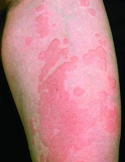
Diseases with fever and rash may be classified by type of eruption: - Centrally distributed maculopapular - Peripheral - Confluent desquamative erythematous - Vesiculobullous - Urticaria-like - Nodular - Purpuric - Ulcerated - With eschars
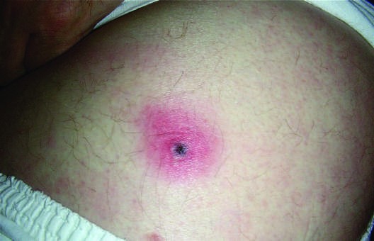
Lesion Morphology Definitions: - Macules: Flat lesions defined by an area of changed color (i.e., a blanchable erythema). - Papules: Raised, solid lesions 5 mm in diameter with a flat, plateau-like surface. - Nodules: Lesions >5 mm in diameter with a more rounded configuration. - Wheals (urticaria, hives): Papules or plaques that are pale pink and may appear annular (ring-like) as they enlarge; classic (nonvasculitic) wheals are transient, lasting only 24 h in any defined area. - Vesicles: 5 mm. - Pustules: Raised lesions containing purulent exudate. - Ulcer: Defect in the skin extending at least into the upper layer of the dermis. - Eschar (tâche noire): Necrotic lesion covered with a black crust. - Palpable purpura: Raised lesion that is due to inflammation of the vessel wall (vasculitis) with subsequent hemorrhage. - Petechiae: Purpuric lesions 3 mm in diameter.
1.2 Approach to the Patient
A thorough history of patients with fever and rash includes the following relevant information: - Immune status - Medications taken within the previous month - Specific travel history - Immunization status - Exposure to domestic pets and other animals - History of animal (including arthropod) bites - Recent dietary exposures - Existence of cardiac abnormalities - Presence of prosthetic material - Recent exposure to ill individuals - Sexual exposures - Site of onset of the rash and its direction and rate of spread
Physical Examination: - Close attention to the rash - Assessment and precise identification of its salient features - Determination of lesion type (macules, papules, etc.) - Distribution (central or peripheral) - Configuration (annular or target) - Arrangement of lesions
2. EPIDEMIOLOGY
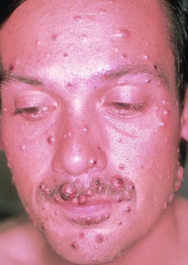
Diseases with fever and rash vary by age, geography, and risk factors. - Measles: Nonimmune individuals. - Rubella: Nonimmune individuals; exposure of pregnant women should be avoided as rubella causes severe congenital abnormalities. - Erythema infectiosum: Most common among children 3–12 years old; occurs in winter and spring. - Exanthem subitum (roseola): Usually affects children <3 years old. - Primary HIV infection: Individuals recently infected with HIV. - Infectious mononucleosis: Adolescents, young adults. - Drug-induced eruption: Variable findings; occurs in previously sensitized individuals. - Epidemic typhus: Exposure to body lice. - Endemic (murine) typhus: Exposure to rat or cat fleas. - Scrub typhus: Endemic in South Pacific, Australia, Asia; transmitted by mites. - Rickettsial spotted fevers: Exposure to ticks; R. conorii in Mediterranean region, India, Africa; R. australis in Australia; R. sibirica in Siberia, Mongolia; R. africae in Africa, Caribbean. - Human monocytotropic ehrlichiosis: Tick-borne; most common in U.S. Southeast, southern Midwest, and mid-Atlantic regions. - Leptospirosis: Exposure to water contaminated with animal urine. - Lyme disease: Bite of Ixodes tick vector. - Southern tick-associated rash illness (STARI): Bite of tick vector Amblyomma americanum (Lone Star tick); often found in regions where Lyme disease is uncommon, including southern United States. - Typhoid fever: Ingestion of contaminated food or water (rare in U.S.). - Dengue fever: Occurs in tropics and subtropics; transmitted by mosquito. - Rat-bite fever: Rat bite; primarily found in Asia; rare in U.S. - Relapsing fever: Exposure to ticks or body lice. - Systemic lupus erythematosus (SLE): Most common in young to middle-aged women; flares precipitated by sun exposure. - Still's disease: Children and young adults. - African trypanosomiasis: Tsetse fly bite in eastern or western Africa. - Arcanobacterial pharyngitis: Children and young adults. - West Nile virus infection: Mosquito bite; rarely, blood transfusion or transplanted organ. - Zika virus infection: Mosquito bite; sexual transmission or blood transfusion less common. - Chronic meningococcemia: Refer to Chapters 160, 161, 202. - Disseminated gonococcal infection: Refer to Chapter 160, 161, 202. - Human parvovirus B19 infection: Refer to Chapter 202. - RIME: Refer to Chapter 160, 161, 202. - Rocky Mountain spotted fever: Tick vector; widespread but more common in southeastern and southwest-central U.S. - Secondary syphilis: Sexually transmitted. - Chikungunya fever: Aedes aegypti and A. albopictus mosquito bites; tropical and subtropical regions. - Hand-foot-and-mouth disease: Summer and fall; primarily children <10 years old; multiple family members. - Erythema multiforme: Herpes simplex virus or Mycoplasma pneumoniae infection; drug intake (i.e., sulfa, phenytoin, penicillin). - Rat-bite fever (Haverhill fever): Rat bite, ingestion of contaminated food. - Bacterial endocarditis: Abnormal heart valve (e.g., viridans streptococci), intravenous drug use. - COVID-19: Infection with SARS-CoV-2. - Multisystem inflammatory syndrome in children (MIS-C): Infection with SARS-CoV-2; MIS-C in older children/adolescents. - Scarlet fever: Most common among children 2–10 years old; usually follows group A streptococcal pharyngitis. - Kawasaki disease: Children <8 years old. - Streptococcal toxic shock syndrome: May occur in setting of severe group A streptococcal infections (e.g., necrotizing fasciitis, bacteremia, pneumonia). - Staphylococcal toxic shock syndrome: Colonization with toxin-producing S. aureus. - Staphylococcal scalded-skin syndrome: Colonization with toxin-producing S. aureus; occurs in children <10 years old (termed Ritter's disease in neonates) or adults with renal dysfunction. - Exfoliative erythroderma: Usually occurs in adults over age 50; more common among men. - DRESS: Individuals genetically unable to detoxify arene oxides (especially hepatic), arene oxide metabolites, lymphocytes; HHV6 viremia. - Stevens-Johnson syndrome (SJS), toxic epidermal necrolysis (TEN): Uncommon among children; more common among people living with HIV, systemic lupus erythematosus, certain HLA types, or slow acetylators. - Varicella: Usually affects children; 10% of adults susceptible; more common in late winter and spring. - Pseudomonas folliculitis: Bathers in hot tubs or swimming pools. - Variola (smallpox): Nonimmune individuals exposed to smallpox.
Figures & Illustrations
Reproduced from Harrison's 22nd Edition.
Figure 1
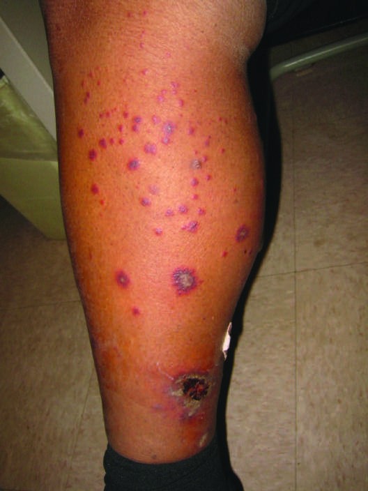
Caption: morphologies, including petechial. Purpuric nodules may develop on FIGURE 21-7 Purpuric lesions of cutaneous small vessel vasculitis in a patient the legs and resemble erythema nodosum but lack its exquisite tender- with Henoch-Schonlein purpura. (Courtesy of Peter Lio, MD; with permission) ness. Lesions of disseminated gonococcemia (Chap. 161) are distinctive, sparse, countable hemorrhagic pustules (Fig. A1-43), usually located distribution. Viral hemorrhagic fever (Chaps. 215 and 216) should be near joints. The lesions of chronic meningococcemia and those of considered in patients with an appropriate travel history and a pete- gonococcemia may be indistinguishable in terms of appearance and chial rash. Thrombotic thrombocytopenic purpura (Chaps. 61, 105, and 120) and hemolytic-uremic syndrome (Chaps. 120, 166, and 172) are closely related and are noninfectious causes of fever and petechiae. Cutaneous small-vessel vasculitis (leukocytoclastic vasculitis) typically — Figure 21-1: Distribution of rubeola (measles) rash starting at the hairline and moving down the body, typically sparing the palms and soles.
Figure 2

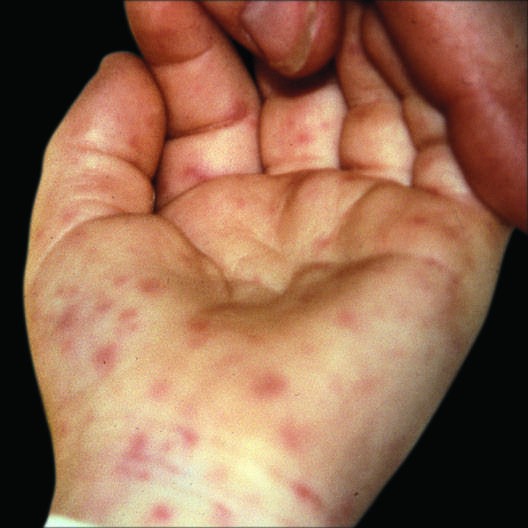
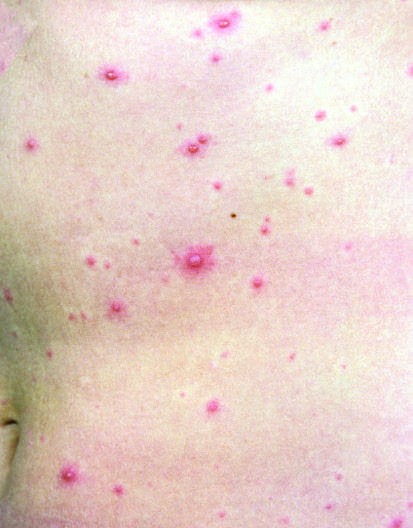
Caption: FIGURE 21-2 Peripheral eruption on the wrist and palm exhibiting erythematous macules in the process of evolving into petechial lesions in a patient with Rocky FIGURE 21-1 Centrally distributed, maculopapular eruption on the trunk in a Mountain spotted fever. (From K Wolff et al [eds]: Fitzpatrick’s Color Atlas and patient with measles. (From EJ Mayeaux Jr et al: Measles, in Usatine RP et al [eds]: Synopsis of Clinical Dermatology, 8th ed. New York, McGraw-Hill, 2017, p. 562, Figure 25-50; with permission.) Color Atlas and Synopsis of Family Medicine, 3rd ed. New York, McGraw-Hill, 2019, p. 797, Figure 132-2. Reproduced with permission from Richard P. Usatine, MD.) Chikungunya fever (Chap. 215), which is transmitted by mosquito bite in tropical and subtropical regions, is associated with a maculopapular (Chap. 191), typically manifests as single or multiple annular lesions. eruption (Fig. A1-54) and severe polyarticular small-joint arthralgias. — Figure 21-2: Rocky Mountain spotted fever rash beginning on wrists and ankles and spreading centripetally to involve palms and soles later in disease.
Figure 3
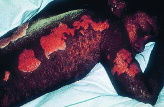
Caption: FIGURE 21-2 Peripheral eruption on the wrist and palm exhibiting erythematous macules in the process of evolving into petechial lesions in a patient with Rocky FIGURE 21-1 Centrally distributed, maculopapular eruption on the trunk in a Mountain spotted fever. (From K Wolff et al [eds]: Fitzpatrick’s Color Atlas and patient with measles. (From EJ Mayeaux Jr et al: Measles, in Usatine RP et al [eds]: Synopsis of Clinical Dermatology, 8th ed. New York, McGraw-Hill, 2017, p. 562, Figure 25-50; with permission.) Color Atlas and Synopsis of Family Medicine, 3rd ed. New York, McGraw-Hill, 2019, p. 797, Figure 132-2. Reproduced with permission from Richard P. Usatine, MD.) Chikungunya fever (Chap. 215), which is transmitted by mosquito bite in tropical and subtropical regions, is associated with a maculopapular (Chap. 191), typically manifests as single or multiple annular lesions. eruption (Fig. A1-54) and severe polyarticular small-joint arthralgias. — Figure 21-3: Stevens-Johnson syndrome (SJS) and toxic epidermal necrolysis (TEN) showing erythematous and purpuric macules progressing to bullae and sloughing of epidermis.
Figure 4
Caption: and Rash FIGURE 21-3 Confluent desquamation in a patient with toxic epidermal necrolysis. fever and toxicity, while recurrent disease (Fig. A1-58C) is milder. (From KS-M Kane et al: Color Atlas & Synopsis of Pediatric Dermatology, 3rd ed. Rickettsialpox (Chap. 192) is often documented in urban settings New York, McGraw Hill, 2017, Figure 15-6; with permission.) and is characterized by vesicles followed by pustules (Figs. A1-33B, A1-33C). It can be distinguished from varicella by an eschar at the site does not exhibit a strawberry tongue or circumoral pallor. In contrast of the mouse-mite bite (Fig. A1-33A) and the papule/plaque base of to the staphylococcal scalded-skin syndrome, in which the cleavage each vesicle. Acute generalized exanthematous pustulosis (Fig. A1-49) plane is superficial in the epidermis, toxic epidermal necrolysis (Chap. 63), should be considered in individuals who are acutely febrile and are a maximal variant of Stevens-Johnson syndrome, involves sloughing taking new medications, especially anticonvulsant or antimicrobial of the entire epidermis (Fig. 21-3, see also Fig. A1-26), resulting in agents (Chap. 63). Disseminated Vibrio vulnificus infection (Chap. 173) — Figure A1-1: Erythema infectiosum (fifth disease) showing bright-red 'slapped-cheeks' appearance followed by lacy reticular rash.
Figure 5
Caption: and Rash FIGURE 21-3 Confluent desquamation in a patient with toxic epidermal necrolysis. fever and toxicity, while recurrent disease (Fig. A1-58C) is milder. (From KS-M Kane et al: Color Atlas & Synopsis of Pediatric Dermatology, 3rd ed. Rickettsialpox (Chap. 192) is often documented in urban settings New York, McGraw Hill, 2017, Figure 15-6; with permission.) and is characterized by vesicles followed by pustules (Figs. A1-33B, A1-33C). It can be distinguished from varicella by an eschar at the site does not exhibit a strawberry tongue or circumoral pallor. In contrast of the mouse-mite bite (Fig. A1-33A) and the papule/plaque base of to the staphylococcal scalded-skin syndrome, in which the cleavage each vesicle. Acute generalized exanthematous pustulosis (Fig. A1-49) plane is superficial in the epidermis, toxic epidermal necrolysis (Chap. 63), should be considered in individuals who are acutely febrile and are a maximal variant of Stevens-Johnson syndrome, involves sloughing taking new medications, especially anticonvulsant or antimicrobial of the entire epidermis (Fig. 21-3, see also Fig. A1-26), resulting in agents (Chap. 63). Disseminated Vibrio vulnificus infection (Chap. 173) — Figure A1-2: Koplik's spots on buccal mucosa, pathognomonic for measles.
Figure 6
Caption: morphologies, including petechial. Purpuric nodules may develop on FIGURE 21-7 Purpuric lesions of cutaneous small vessel vasculitis in a patient the legs and resemble erythema nodosum but lack its exquisite tender- with Henoch-Schonlein purpura. (Courtesy of Peter Lio, MD; with permission) ness. Lesions of disseminated gonococcemia (Chap. 161) are distinctive, sparse, countable hemorrhagic pustules (Fig. A1-43), usually located distribution. Viral hemorrhagic fever (Chaps. 215 and 216) should be near joints. The lesions of chronic meningococcemia and those of considered in patients with an appropriate travel history and a pete- gonococcemia may be indistinguishable in terms of appearance and chial rash. Thrombotic thrombocytopenic purpura (Chaps. 61, 105, and 120) and hemolytic-uremic syndrome (Chaps. 120, 166, and 172) are closely related and are noninfectious causes of fever and petechiae. Cutaneous small-vessel vasculitis (leukocytoclastic vasculitis) typically — Figure A1-8: Lyme disease showing erythema migrans papule expanding to erythematous annular lesion with central clearing.
Figure 7
Caption: and Rash FIGURE 21-3 Confluent desquamation in a patient with toxic epidermal necrolysis. fever and toxicity, while recurrent disease (Fig. A1-58C) is milder. (From KS-M Kane et al: Color Atlas & Synopsis of Pediatric Dermatology, 3rd ed. Rickettsialpox (Chap. 192) is often documented in urban settings New York, McGraw Hill, 2017, Figure 15-6; with permission.) and is characterized by vesicles followed by pustules (Figs. A1-33B, A1-33C). It can be distinguished from varicella by an eschar at the site does not exhibit a strawberry tongue or circumoral pallor. In contrast of the mouse-mite bite (Fig. A1-33A) and the papule/plaque base of to the staphylococcal scalded-skin syndrome, in which the cleavage each vesicle. Acute generalized exanthematous pustulosis (Fig. A1-49) plane is superficial in the epidermis, toxic epidermal necrolysis (Chap. 63), should be considered in individuals who are acutely febrile and are a maximal variant of Stevens-Johnson syndrome, involves sloughing taking new medications, especially anticonvulsant or antimicrobial of the entire epidermis (Fig. 21-3, see also Fig. A1-26), resulting in agents (Chap. 63). Disseminated Vibrio vulnificus infection (Chap. 173) — Figure A1-23: Bacterial endocarditis showing Osler's nodes (tender pink nodules on finger or toe pads) and Janeway lesions (painless erythematous macules on palms and soles).
Figure 8
Caption: morphologies, including petechial. Purpuric nodules may develop on FIGURE 21-7 Purpuric lesions of cutaneous small vessel vasculitis in a patient the legs and resemble erythema nodosum but lack its exquisite tender- with Henoch-Schonlein purpura. (Courtesy of Peter Lio, MD; with permission) ness. Lesions of disseminated gonococcemia (Chap. 161) are distinctive, sparse, countable hemorrhagic pustules (Fig. A1-43), usually located distribution. Viral hemorrhagic fever (Chaps. 215 and 216) should be near joints. The lesions of chronic meningococcemia and those of considered in patients with an appropriate travel history and a pete- gonococcemia may be indistinguishable in terms of appearance and chial rash. Thrombotic thrombocytopenic purpura (Chaps. 61, 105, and 120) and hemolytic-uremic syndrome (Chaps. 120, 166, and 172) are closely related and are noninfectious causes of fever and petechiae. Cutaneous small-vessel vasculitis (leukocytoclastic vasculitis) typically — Figure A1-29: Kawasaki disease showing rash similar to scarlet fever, strawberry tongue, and edema of hands and feet.
Generated from Harrison's Principles of Internal Medicine, 22nd Edition.