Interventional Nephrology
Chapter 324 | Part 9: Disorders of the Kidney and Urinary Tract · Part 9 – Renal & Urinary Tract Disorders
Detailed clinical reference synthesised from Harrison's Principles of Internal Medicine, 22nd Edition
🔑 Key Clinical Points
- Interventional nephrology focuses on dialysis access for peritoneal and hemodialysis, typically performed under fluoroscopy.
- Fistula First and Catheter Last campaigns have reduced arteriovenous graft (AVG) prevalence to <20% in the US, increasing autogenous arteriovenous fistula (AVF) prevalence to near 65%.
- High-access flow (>1500 mL/min) can lead to systemic complications like heart failure and pulmonary hypertension.
- Physical examination of dialysis access involves assessing pulsatility, murmurs, thrill, augmentation, and collapse to detect inflow/outflow stenosis.
- Encapsulating peritoneal sclerosis is a late-stage complication of peritoneal dialysis triggered by repeated peritonitis.
- Preservation of venous real estate is critical, especially for patients needing cardiac rhythm management devices (CRMDs).
- Banding (using a 2-0 Prolene suture) is a common procedure to reduce access flows and prevent steal syndrome.
- Catheter-related bacteremia is best treated with exchange of the catheter and disruption of any fibrinous sheath.
- Steal syndrome typically presents as ischemia of the hand due to high-access flow.
- Omental entrapment of the peritoneal catheter often requires laparoscopic intervention or omentopexy at placement.
📑 Table of Contents
- 1. DEFINITION & OVERVIEW
- 1.1 History of Vascular Access
- 2. EPIDEMIOLOGY
- 2.1 Access Utilization Trends
- 3. ETIOLOGY & PATHOPHYSIOLOGY
- 3.1 Peritoneal Dialysis Catheter Pathophysiology
- 3.2 Hemodialysis Catheter Pathophysiology
- 3.3 Arteriovenous Fistula and Graft Pathophysiology
- 4. CLINICAL FEATURES
- 4.1 Physical Examination of Dialysis Access
- 5. DIFFERENTIAL DIAGNOSIS
- 5.1 Stenosis Differentiation
- 6. INVESTIGATIONS & DIAGNOSIS
- 6.1 Physical Examination Findings
- 7. MANAGEMENT & TREATMENT
- 7.1 Catheter Management
- 7.2 Access Flow Reduction
- 7.3 Peritoneal Dialysis Complications
- 8. PROGNOSIS & COMPLICATIONS
- 8.1 Systemic Complications
- 8.2 Skin and Access Breakdown
- 8.3 Infection and Thrombosis
- 9. SPECIAL CONSIDERATIONS
- 9.1 Transplant Access
- 9.2 Cardiac Devices
- 9.3 Hospitalization Access
- 10. KEY PEARLS & CLINICAL TRAPS
- 10.1 Clinical Pearls
- Figures & Illustrations
📋 Figures in This Chapter
1. DEFINITION & OVERVIEW
- Interventional nephrology is a procedure-oriented subspecialty with a focus on dialysis access for peritoneal and hemodialysis, typically performed under fluoroscopy.
- Interventional nephrologists (INs) usually provide patient care in multidisciplinary teams that include clinical nephrologists; access surgeons with vascular, transplant, or general surgery background; other interventionists (with radiology or cardiology training); and dialysis unit access coordinators, nurses, and technicians involved in needle replacement.
- Long-term preservation of venous and arterial vascular access options is one tenet of chronic kidney disease (CKD) care, leading INs to advocate for specific vascular access options (tunneled small-diameter catheters over peripherally inserted central catheters [PICCs] and cardiac devices [epicardial rather than endovascular lead passage]).
- The history of vascular access for hemodialysis is closely tied to the history of dialysis.
- The first hemodialysis treatments in humans were performed in 1924 using glass needles to access the radial artery and return blood into the cubital vein.
- In 1943, a 'rotating drum kidney' was used to dialyze a 29-year-old housemaid with CKD by surgical access.
- Catheter-based approaches for chronic renal replacement therapy (RRT) were designed initially in 1961 for hemodialysis and in 1968 for peritoneal dialysis, both using Dacron felt cuffs to protect against infection.
- Material sciences have continued to evolve the development of grafts for use in hemodialysis: A modified bovine carotid artery biological graft was introduced in 1972, followed by the use of expanded polytetrafluoroethylene (ePTFE) grafts in 1976, and most recently in 2016, tissue-engineered blood vessels from human fibroblasts and endothelial cells.
- Some ePTFE grafts are modified with a silicone layer to allow for early cannulation within days of insertion.
- Ultra-high-pressure (up to 40 atm) angioplasty balloons are a mainstay of peripheral and central venous therapy, and Nitinol self-expanding stents and stent grafts serve as rescue tools for unsuccessful angioplasty as well as vessel rupture with extravasation.
- The role of drug-coated balloons in dialysis access care continues to be assessed, although greater cost and, to date, mixed clinical trial results have limited widespread use.
1.1 History of Vascular Access
- The first hemodialysis treatments in humans were performed in 1924 using glass needles to access the radial artery and return blood into the cubital vein.
- In 1943, a 'rotating drum kidney' was used to dialyze a 29-year-old housemaid with CKD by surgical access.
- Catheter-based approaches for chronic renal replacement therapy (RRT) were designed initially in 1961 for hemodialysis and in 1968 for peritoneal dialysis, both using Dacron felt cuffs to protect against infection.
- Material sciences have continued to evolve the development of grafts for use in hemodialysis: A modified bovine carotid artery biological graft was introduced in 1972, followed by the use of expanded polytetrafluoroethylene (ePTFE) grafts in 1976, and most recently in 2016, tissue-engineered blood vessels from human fibroblasts and endothelial cells.
- Some ePTFE grafts are modified with a silicone layer to allow for early cannulation within days of insertion.
- Ultra-high-pressure (up to 40 atm) angioplasty balloons are a mainstay of peripheral and central venous therapy, and Nitinol self-expanding stents and stent grafts serve as rescue tools for unsuccessful angioplasty as well as vessel rupture with extravasation.
- The role of drug-coated balloons in dialysis access care continues to be assessed, although greater cost and, to date, mixed clinical trial results have limited widespread use.
2. EPIDEMIOLOGY
- In the United States, the United States was an international outlier in the use of nonautogenous accesses (arteriovenous grafts [AVGs]).
- In the mid-1990s, 65% of prevalent dialysis patients used an AVG for access.
- Studies associated increased mortality in U.S. dialysis patients with lower AVF prevalence.
- In the context of 'Fistula First' and 'Fistula First, Catheter Last' campaigns, AVG prevalence decreased to its current value of <20%, while AVFs increased to near 65% prevalence.
- However, most centers still struggle with the challenging conditions of arteries and veins in these patients requiring that 75% of AVFs are now created in the upper arm, where the veins a priori are larger in diameter, and arteries can deliver higher blood flow rates due to large vessel diameter.
- Approximately 8–10% of the CKD population has the need for cardiac rhythm management devices (CRMDs) that can lead to loss of the upper arm cephalic vein as well as central venous stenoses and occlusions around device leads.
- CKD patients also have an increased frequency of hospitalizations, some of which require intravenous access beyond the hospital stay for antibiotics, nutritional support, or hydration.
- Avoiding PICCs in a patient with CKD stage 3 or 3b and higher and, instead, using internal (or external) jugular vein tunneled small-diameter catheters preserves arm veins for long-term access creation.
2.1 Access Utilization Trends
- In the United States, the United States was an international outlier in the use of nonautogenous accesses (arteriovenous grafts [AVGs]).
- In the mid-1990s, 65% of prevalent dialysis patients used an AVG for access.
- Studies associated increased mortality in U.S. dialysis patients with lower AVF prevalence.
- In the context of 'Fistula First' and 'Fistula First, Catheter Last' campaigns, AVG prevalence decreased to its current value of <20%, while AVFs increased to near 65% prevalence.
- However, most centers still struggle with the challenging conditions of arteries and veins in these patients requiring that 75% of AVFs are now created in the upper arm, where the veins a priori are larger in diameter, and arteries can deliver higher blood flow rates due to large vessel diameter.
3. ETIOLOGY & PATHOPHYSIOLOGY
- Peritoneal dialysis (PD) catheters can be placed fluoroscopically, peritoneoscopically, laparoscopically, and open surgically.
- Procedural success is typically linked to provider experience and procedural planning to optimize positioning of the PD catheter coil as this improves function and decreases drain pain and other complications.
- The internal cuff is placed within the rectus sheath just laterally to the linea alba, while the external PD catheter cuff should be located 2–4 cm from the skin exit site.
- Ingrowth of both cuffs ensures secure positioning of the catheter and allows water emersion.
- Over time, the peritoneal catheter can become encased in a fibrinous sheath, which, if limiting fluid flow during exchanges, can be disrupted by guidewire manipulation.
- Omental entrapment of the catheter often requires laparoscopic intervention; omentopexy at the time of PD catheter placement can prevent later entrapment.
- Repeated infections affect the permeability of the peritoneal membrane, as does long-term exposure to glucose-containing exchange solutions.
- Encapsulating peritoneal sclerosis is a late-stage complication of PD thought to be triggered by repeated peritonitis.
- Dialysis catheters are typically made of polyurethane that softens at body temperature but is sufficiently strong to allow for blood flow rates of 400–500 mL/min in each of two channels inside a 14.5–16 French design without collapse of the catheter lumen.
- Tunneled catheters have a cuff that creates a barrier between skin flora at the exit site and the sterile catheter tunnel leading into the fibrinous sheath covering the catheter from the vessel insertion point to its tip.
- The fibrinous sheath can extend too far, impeding catheter flow and necessitating exchange of the catheter with disruption of the sheath by balloon angioplasty.
- Catheter-related bacteremia is best treated with exchange of the catheter and disruption of any fibrinous sheath, although removal of the catheter and delayed reinsertion after several days is also successful.
- Catheter infection-related sepsis unresponsive to antibiotics requires removal of the catheter.
- Thrombotic occlusion and later sclerotic scarring of the vein at catheter insertion sites is common, however, and removal of a catheter may lead to loss of this access site.
- Catheter vessel wall contact points are thought to lead to central vein stenosis, which is more commonly observed in patients with catheter contact times of longer duration (>3 months).
- Catheter tip position in the large central veins instead of the right atrium causes additional injury from dynamic blood movement during dialysis treatments and should be corrected.
- A thrombus is commonly found attached to the catheter, often tethering the catheter to the vessel wall and right atrium.
- While some thrombi are mobile and dissolve with anticoagulation, a wall-tethered thrombus is often well organized with cellular components and quite resistant to pharmacologic lysis.
- Clinically significant pulmonary embolism from catheter-associated thrombus is rare, and it may be that only intra-atrial thrombus >2 cm in diameter deserves active intervention.
- To provide successful dialysis, an AVF or AVG has to provide at least the desired blood pump speed plus 100–200 mL/min to minimize recirculation and prevent collapse of the access.
- In the United States, this usually means flow in the 600–800 mL/min range.
- A fistula created using an artery with ≥2 mm diameter and a vein ≥3 mm will typically have flow volumes >500 mL/min when the systolic blood pressure is >100 mmHg.
- After creation of the arterial-venous anastomosis (or insertion of the AVG), blood flow increases significantly: brachial artery flow at rest is typically 10 mm in diameter in the upper arm such that the artery continues to enlarge until a narrow segment in the venous conduit becomes flow limiting.
- Flow volumes in these mature upper arm AVFs are usually 1400–1800 mL/min, but after a few years can be as high as 2000–4000 mL/min.
- Forearm AVFs usually have lower flow volumes (500–700 mL/min) as the feeding radial artery is of smaller diameter and, in the context of systemic vascular disease in the United States, only increases in diameter over many years.
- Increased flows and pressure in the venous segment of the access circuit combine to lead to 'chronic dialysis access disease' that manifests differently for each type of the common long-term accesses in predetermined segments particularly prone to shear stress and needle insertion–related injury.
- AVGs develop venous anastomotic stenoses that recur with very short periodicity in the 3- to 4-month range.
- Stent grafts can effectively be deployed to extend patency for usually 1 year at the site, after which the buildup of pauci-cellular fibrous depositions and then at the stent edges requires re-angioplasty one to three times per year.
- Forearm radial-cephalic autogenous accesses of the Cimino type at the wrist are most prone to low flow due to juxta-anastomotic stenoses.
- Over time, these stenoses can stabilize, and with enlargement of the inflow artery, they effectively provide protection against excessive flows and their sequelae.
- 'Snuff box' radial-cephalic fistulas require minimal mobilization of the cephalic vein with fewer juxta-anastomotic stenoses.
- However, the additional side branches and associated venous valves may present as stenosis and require ligation to allow cannulation.
- Upper arm brachial-cephalic autogenous accesses typically develop stenoses in the cephalic arch, which recur in accelerated fashion after each angioplasty.
- Flexible stent grafts in the cephalic arch extend intraprocedural intervals usually to 9–12 months.
- Upper arm transposed brachial-basilic autogenous accesses develop stenoses in the swing point where the basilic vein is curved during a mobilization procedure to provide a location more lateral and closer to the skin to facilitate needle cannulation.
- Angioplasty and stent graft placement approaches extend patency.
- In both types of upper arm accesses, there are often prolonged periods with increased intra-access pressures due to outflow stenoses, which lead to enlargement of needle insertion site aneurysm as the skin heals in a pressurized, stretched state.
- Continued use of pressurized accesses leads to enlargement of needle sites, then thinning of the skin, scab formation, and, finally, full-thickness ulceration with often significant bleeding events.
- Recognizing the occurrence of outflow stenoses early is an important skill for nurses and technologists working in dialysis units to acquire in order to avoid irreversible loss of skin coverage and possible loss of the access.
- High-access flow (>1500 mL/min) can lead to systemic complications, such as heart failure and pulmonary hypertension.
- Fistula inflow higher than outflow capacity leads to accelerated aneurysm formation and breakdown of skin coverage as intra-access pressures are increased over the ideal pressure of 20–35 mmHg.
- High-access flows are also associated with steal syndrome, typically ischemia of the hand.
- A variety of procedures have been described to reduce access flows, the most common being 'banding,' where typically a 2-0 Prolene suture is guided around the inflow and a 3- or 4-mm spacer and is tied snugly over the spacer to create an inflow stenosis.
- An outflow stenosis will decrease the degree of collapse; banding of an upper arm access or a natural flow-limiting stenosis may lead to complete collapse of an upper arm access.
- Enlarged needle insertion sites (and any sites of suspected skin thinning) are best examined while occluding inflow: the completely empty access allows palpation of a firm, layered thrombus inside aneurysms as well as a better appreciation of the thickness of the overlying skin by rolling it between thumb and index finger.
- The chest wall and neck should be inspected for the presence of skin veins and venous distention, which are associated with central venous stenosis or occlusion, as is ipsilateral arm edema.
3.1 Peritoneal Dialysis Catheter Pathophysiology
- Peritoneal dialysis (PD) catheters can be placed fluoroscopically, peritoneoscopically, laparoscopically, and open surgically.
- Procedural success is typically linked to provider experience and procedural planning to optimize positioning of the PD catheter coil as this improves function and decreases drain pain and other complications.
- The internal cuff is placed within the rectus sheath just laterally to the linea alba, while the external PD catheter cuff should be located 2–4 cm from the skin exit site.
- Ingrowth of both cuffs ensures secure positioning of the catheter and allows water emersion.
- Over time, the peritoneal catheter can become encased in a fibrinous sheath, which, if limiting fluid flow during exchanges, can be disrupted by guidewire manipulation.
- Omental entrapment of the catheter often requires laparoscopic intervention; omentopexy at the time of PD catheter placement can prevent later entrapment.
- Repeated infections affect the permeability of the peritoneal membrane, as does long-term exposure to glucose-containing exchange solutions.
- Encapsulating peritoneal sclerosis is a late-stage complication of PD thought to be triggered by repeated peritonitis.
3.2 Hemodialysis Catheter Pathophysiology
- Dialysis catheters are typically made of polyurethane that softens at body temperature but is sufficiently strong to allow for blood flow rates of 400–500 mL/min in each of two channels inside a 14.5–16 French design without collapse of the catheter lumen.
- Tunneled catheters have a cuff that creates a barrier between skin flora at the exit site and the sterile catheter tunnel leading into the fibrinous sheath covering the catheter from the vessel insertion point to its tip.
- The fibrinous sheath can extend too far, impeding catheter flow and necessitating exchange of the catheter with disruption of the sheath by balloon angioplasty.
- Catheter-related bacteremia is best treated with exchange of the catheter and disruption of any fibrinous sheath, although removal of the catheter and delayed reinsertion after several days is also successful.
- Catheter infection-related sepsis unresponsive to antibiotics requires removal of the catheter.
- Thrombotic occlusion and later sclerotic scarring of the vein at catheter insertion sites is common, however, and removal of a catheter may lead to loss of this access site.
- Catheter vessel wall contact points are thought to lead to central vein stenosis, which is more commonly observed in patients with catheter contact times of longer duration (>3 months).
- Catheter tip position in the large central veins instead of the right atrium causes additional injury from dynamic blood movement during dialysis treatments and should be corrected.
- A thrombus is commonly found attached to the catheter, often tethering the catheter to the vessel wall and right atrium.
- While some thrombi are mobile and dissolve with anticoagulation, a wall-tethered thrombus is often well organized with cellular components and quite resistant to pharmacologic lysis.
- Clinically significant pulmonary embolism from catheter-associated thrombus is rare, and it may be that only intra-atrial thrombus >2 cm in diameter deserves active intervention.
3.3 Arteriovenous Fistula and Graft Pathophysiology
- To provide successful dialysis, an AVF or AVG has to provide at least the desired blood pump speed plus 100–200 mL/min to minimize recirculation and prevent collapse of the access.
- In the United States, this usually means flow in the 600–800 mL/min range.
- A fistula created using an artery with ≥2 mm diameter and a vein ≥3 mm will typically have flow volumes >500 mL/min when the systolic blood pressure is >100 mmHg.
- After creation of the arterial-venous anastomosis (or insertion of the AVG), blood flow increases significantly: brachial artery flow at rest is typically 10 mm in diameter in the upper arm such that the artery continues to enlarge until a narrow segment in the venous conduit becomes flow limiting.
- Flow volumes in these mature upper arm AVFs are usually 1400–1800 mL/min, but after a few years can be as high as 2000–4000 mL/min.
- Forearm AVFs usually have lower flow volumes (500–700 mL/min) as the feeding radial artery is of smaller diameter and, in the context of systemic vascular disease in the United States, only increases in diameter over many years.
- Increased flows and pressure in the venous segment of the access circuit combine to lead to 'chronic dialysis access disease' that manifests differently for each type of the common long-term accesses in predetermined segments particularly prone to shear stress and needle insertion–related injury.
- AVGs develop venous anastomotic stenoses that recur with very short periodicity in the 3- to 4-month range.
- Stent grafts can effectively be deployed to extend patency for usually 1 year at the site, after which the buildup of pauci-cellular fibrous depositions and then at the stent edges requires re-angioplasty one to three times per year.
- Forearm radial-cephalic autogenous accesses of the Cimino type at the wrist are most prone to low flow due to juxta-anastomotic stenoses.
- Over time, these stenoses can stabilize, and with enlargement of the inflow artery, they effectively provide protection against excessive flows and their sequelae.
- 'Snuff box' radial-cephalic fistulas require minimal mobilization of the cephalic vein with fewer juxta-anastomotic stenoses.
- However, the additional side branches and associated venous valves may present as stenosis and require ligation to allow cannulation.
- Upper arm brachial-cephalic autogenous accesses typically develop stenoses in the cephalic arch, which recur in accelerated fashion after each angioplasty.
- Flexible stent grafts in the cephalic arch extend intraprocedural intervals usually to 9–12 months.
- Upper arm transposed brachial-basilic autogenous accesses develop stenoses in the swing point where the basilic vein is curved during a mobilization procedure to provide a location more lateral and closer to the skin to facilitate needle cannulation.
- Angioplasty and stent graft placement approaches extend patency.
- In both types of upper arm accesses, there are often prolonged periods with increased intra-access pressures due to outflow stenoses, which lead to enlargement of needle insertion site aneurysm as the skin heals in a pressurized, stretched state.
- Continued use of pressurized accesses leads to enlargement of needle sites, then thinning of the skin, scab formation, and, finally, full-thickness ulceration with often significant bleeding events.
- Recognizing the occurrence of outflow stenoses early is an important skill for nurses and technologists working in dialysis units to acquire in order to avoid irreversible loss of skin coverage and possible loss of the access.
- High-access flow (>1500 mL/min) can lead to systemic complications, such as heart failure and pulmonary hypertension.
- Fistula inflow higher than outflow capacity leads to accelerated aneurysm formation and breakdown of skin coverage as intra-access pressures are increased over the ideal pressure of 20–35 mmHg.
- High-access flows are also associated with steal syndrome, typically ischemia of the hand.
- A variety of procedures have been described to reduce access flows, the most common being 'banding,' where typically a 2-0 Prolene suture is guided around the inflow and a 3- or 4-mm spacer and is tied snugly over the spacer to create an inflow stenosis.
- An outflow stenosis will decrease the degree of collapse; banding of an upper arm access or a natural flow-limiting stenosis may lead to complete collapse of an upper arm access.
- Enlarged needle insertion sites (and any sites of suspected skin thinning) are best examined while occluding inflow: the completely empty access allows palpation of a firm, layered thrombus inside aneurysms as well as a better appreciation of the thickness of the overlying skin by rolling it between thumb and index finger.
- The chest wall and neck should be inspected for the presence of skin veins and venous distention, which are associated with central venous stenosis or occlusion, as is ipsilateral arm edema.
4. CLINICAL FEATURES
- The 2019 Kidney Disease Outcomes Quality Initiative (KDOQI) vascular access guidelines were developed under the tenet, '[t]he right access for the right patient at the right time.'
- Progression of CKD is highly variable, many patients die from other causes before reaching end-stage renal disease (ESRD), and some AVFs require 6–12 months to mature to usability in patients with hypertension and diabetes, leading to uncertainty as to when to create AVFs.
- The more common need of upper arm accesses for interventions to maintain patency favors the creation of forearm accesses during the pre-ESRD period.
- The processes of care from vein mapping, surgery, follow-up visits after access creation, to availability and timing of open surgical or endovascular interventions have profound effects on the overall success rate needed to achieve mature and usable accesses and appear to be key factor in the highly variable outcomes across the United States.
- A central skill in dialysis access evaluation is the physical examination.
- Five steps in the access examination capture all aspects of possible pathology: Pulsatility reflects the force of access expansion during systole and the degree of softening during diastole.
- Very high blood pressures will suggest increased pulsatility, but the access softens remarkably during diastole.
- An outflow stenosis will lead to increased pulsatility and reduced softening during diastole.
- An inflow stenosis will blunt the systolic component and create the impression of an 'empty' access during diastole unless there is a coexisting outflow stenosis.
- The audible flow murmur can be characterized by pitch and continuity (Video 324-1).
- A change in pitch toward higher frequency is typical at the site of a stenosis due to accelerated flow velocity at this site.
- A discontinuous flow murmur indicates that during diastole flow is so low that no audible shear force is created; this is the sign of a severe inflow or outflow stenosis.
- Typically, the stenotic inflow murmur is faint (like a whistle), whereas the stenotic outflow murmur can be coarse and loud (akin to a handsaw) (Video 324-2).
- A thrill is palpable through the skin when the vessel is close enough to the surface and the flow high enough in relation to the diameter of the vessel to create vibration of the vessel wall.
- A continuous thrill can be a sign of a well-developed access, usually in the inflow segment, dissipating as the access vessel branches and takes a deeper course.
- In contrast, a discontinuous thrill is found with severe stenosis.
- An isolated thrill is also found focally immediately after a stenosis.
- The differentiation from a 'healthy' thrill can be made by documenting a change in pulsatility at the site of the focal thrill, increased retrograde (inflow) and decreased antegrade (outflow).
- Augmentation is the engorgement of the body of the access (where needles are inserted) with occlusion of the outflow necessary for safe and successful needle insertions.
- An inflow stenosis will impair augmentation, as will side branches of the occluding finger/tourniquet and the inflow.
- The location of side branches can be elucidated by moving the occluding finger closer toward the anastomosis until augmentation is achieved.
- With several collaterals, this may be a staged phenomenon.
- Collapse of the access with arm elevation (against gravity) is a measure of inflow and outflow capacity match or mismatch.
- A forearm access typically displays complete collapse, while upper arm accesses typically show only partial collapse.
- An outflow stenosis or very high inflow will decrease the degree of collapse; banding of an upper arm access or a natural flow-limiting stenosis may lead to complete collapse of an upper arm access.
4.1 Physical Examination of Dialysis Access
- The 2019 Kidney Disease Outcomes Quality Initiative (KDOQI) vascular access guidelines were developed under the tenet, '[t]he right access for the right patient at the right time.'
- Progression of CKD is highly variable, many patients die from other causes before reaching end-stage renal disease (ESRD), and some AVFs require 6–12 months to mature to usability in patients with hypertension and diabetes, leading to uncertainty as to when to create AVFs.
- The more common need of upper arm accesses for interventions to maintain patency favors the creation of forearm accesses during the pre-ESRD period.
- The processes of care from vein mapping, surgery, follow-up visits after access creation, to availability and timing of open surgical or endovascular interventions have profound effects on the overall success rate needed to achieve mature and usable accesses and appear to be key factor in the highly variable outcomes across the United States.
- A central skill in dialysis access evaluation is the physical examination.
- Five steps in the access examination capture all aspects of possible pathology: Pulsatility reflects the force of access expansion during systole and the degree of softening during diastole.
- Very high blood pressures will suggest increased pulsatility, but the access softens remarkably during diastole.
- An outflow stenosis will lead to increased pulsatility and reduced softening during diastole.
- An inflow stenosis will blunt the systolic component and create the impression of an 'empty' access during diastole unless there is a coexisting outflow stenosis.
- The audible flow murmur can be characterized by pitch and continuity (Video 324-1).
- A change in pitch toward higher frequency is typical at the site of a stenosis due to accelerated flow velocity at this site.
- A discontinuous flow murmur indicates that during diastole flow is so low that no audible shear force is created; this is the sign of a severe inflow or outflow stenosis.
- Typically, the stenotic inflow murmur is faint (like a whistle), whereas the stenotic outflow murmur can be coarse and loud (akin to a handsaw) (Video 324-2).
- A thrill is palpable through the skin when the vessel is close enough to the surface and the flow high enough in relation to the diameter of the vessel to create vibration of the vessel wall.
- A continuous thrill can be a sign of a well-developed access, usually in the inflow segment, dissipating as the access vessel branches and takes a deeper course.
- In contrast, a discontinuous thrill is found with severe stenosis.
- An isolated thrill is also found focally immediately after a stenosis.
- The differentiation from a 'healthy' thrill can be made by documenting a change in pulsatility at the site of the focal thrill, increased retrograde (inflow) and decreased antegrade (outflow).
- Augmentation is the engorgement of the body of the access (where needles are inserted) with occlusion of the outflow necessary for safe and successful needle insertions.
- An inflow stenosis will impair augmentation, as will side branches of the occluding finger/tourniquet and the inflow.
- The location of side branches can be elucidated by moving the occluding finger closer toward the anastomosis until augmentation is achieved.
- With several collaterals, this may be a staged phenomenon.
- Collapse of the access with arm elevation (against gravity) is a measure of inflow and outflow capacity match or mismatch.
- A forearm access typically displays complete collapse, while upper arm accesses typically show only partial collapse.
- An outflow stenosis or very high inflow will decrease the degree of collapse; banding of an upper arm access or a natural flow-limiting stenosis may lead to complete collapse of an upper arm access.
5. DIFFERENTIAL DIAGNOSIS
- Differential diagnosis of dialysis access stenosis includes inflow stenosis vs outflow stenosis.
- Inflow stenosis will blunt the systolic component and create the impression of an 'empty' access during diastole unless there is a coexisting outflow stenosis.
- An outflow stenosis will lead to increased pulsatility and reduced softening during diastole.
- Steal syndrome typically presents as ischemia of the hand due to high-access flow.
- Catheter infection-related sepsis unresponsive to antibiotics requires removal of the catheter.
- Encapsulating peritoneal sclerosis is a late-stage complication of PD thought to be triggered by repeated peritonitis.
5.1 Stenosis Differentiation
- Differential diagnosis of dialysis access stenosis includes inflow stenosis vs outflow stenosis.
- Inflow stenosis will blunt the systolic component and create the impression of an 'empty' access during diastole unless there is a coexisting outflow stenosis.
- An outflow stenosis will lead to increased pulsatility and reduced softening during diastole.
6. INVESTIGATIONS & DIAGNOSIS
- Interventional nephrology is a procedure-oriented subspecialty with a focus on dialysis access for peritoneal and hemodialysis, typically performed under fluoroscopy.
- Ultrasound (US) evaluation of dialysis access is common, and some practitioners perform renal and renal artery US evaluation as well as renal biopsies.
- Endovascular creation of arteriovenous fistulas (AVFs) is a recent addition to the procedural spectrum; (open) surgical access creation by nephrologists is limited to very few centers in the United States, while common in China, Germany, India, and Italy.
- The audible flow murmur can be characterized by pitch and continuity (Video 324-1).
- A change in pitch toward higher frequency is typical at the site of a stenosis due to accelerated flow velocity at this site.
- A discontinuous flow murmur indicates that during diastole flow is so low that no audible shear force is created; this is the sign of a severe inflow or outflow stenosis.
- Typically, the stenotic inflow murmur is faint (like a whistle), whereas the stenotic outflow murmur can be coarse and loud (akin to a handsaw) (Video 324-2).
- A thrill is palpable through the skin when the vessel is close enough to the surface and the flow high enough in relation to the diameter of the vessel to create vibration of the vessel wall.
- A continuous thrill can be a sign of a well-developed access, usually in the inflow segment, dissipating as the access vessel branches and takes a deeper course.
- In contrast, a discontinuous thrill is found with severe stenosis.
- An isolated thrill is also found focally immediately after a stenosis.
- The differentiation from a 'healthy' thrill can be made by documenting a change in pulsatility at the site of the focal thrill, increased retrograde (inflow) and decreased antegrade (outflow).
- Augmentation is the engorgement of the body of the access (where needles are inserted) with occlusion of the outflow necessary for safe and successful needle insertions.
- An inflow stenosis will impair augmentation, as will side branches of the occluding finger/tourniquet and the inflow.
- The location of side branches can be elucidated by moving the occluding finger closer toward the anastomosis until augmentation is achieved.
- With several collaterals, this may be a staged phenomenon.
- Collapse of the access with arm elevation (against gravity) is a measure of inflow and outflow capacity match or mismatch.
- A forearm access typically displays complete collapse, while upper arm accesses typically show only partial collapse.
- An outflow stenosis or very high inflow will decrease the degree of collapse; banding of an upper arm access or a natural flow-limiting stenosis may lead to complete collapse of an upper arm access.
6.1 Physical Examination Findings
- The audible flow murmur can be characterized by pitch and continuity (Video 324-1).
- A change in pitch toward higher frequency is typical at the site of a stenosis due to accelerated flow velocity at this site.
- A discontinuous flow murmur indicates that during diastole flow is so low that no audible shear force is created; this is the sign of a severe inflow or outflow stenosis.
- Typically, the stenotic inflow murmur is faint (like a whistle), whereas the stenotic outflow murmur can be coarse and loud (akin to a handsaw) (Video 324-2).
- A thrill is palpable through the skin when the vessel is close enough to the surface and the flow high enough in relation to the diameter of the vessel to create vibration of the vessel wall.
- A continuous thrill can be a sign of a well-developed access, usually in the inflow segment, dissipating as the access vessel branches and takes a deeper course.
- In contrast, a discontinuous thrill is found with severe stenosis.
- An isolated thrill is also found focally immediately after a stenosis.
- The differentiation from a 'healthy' thrill can be made by documenting a change in pulsatility at the site of the focal thrill, increased retrograde (inflow) and decreased antegrade (outflow).
- Augmentation is the engorgement of the body of the access (where needles are inserted) with occlusion of the outflow necessary for safe and successful needle insertions.
- An inflow stenosis will impair augmentation, as will side branches of the occluding finger/tourniquet and the inflow.
- The location of side branches can be elucidated by moving the occluding finger closer toward the anastomosis until augmentation is achieved.
- With several collaterals, this may be a staged phenomenon.
- Collapse of the access with arm elevation (against gravity) is a measure of inflow and outflow capacity match or mismatch.
- A forearm access typically displays complete collapse, while upper arm accesses typically show only partial collapse.
- An outflow stenosis or very high inflow will decrease the degree of collapse; banding of an upper arm access or a natural flow-limiting stenosis may lead to complete collapse of an upper arm access.
7. MANAGEMENT & TREATMENT
- Catheter-related bacteremia is best treated with exchange of the catheter and disruption of any fibrinous sheath, although removal of the catheter and delayed reinsertion after several days is also successful.
- Catheter infection-related sepsis unresponsive to antibiotics requires removal of the catheter.
- Thrombotic occlusion and later sclerotic scarring of the vein at catheter insertion sites is common, however, and removal of a catheter may lead to loss of this access site.
- A variety of procedures have been described to reduce access flows, the most common being 'banding,' where typically a 2-0 Prolene suture is guided around the inflow and a 3- or 4-mm spacer and is tied snugly over the spacer to create an inflow stenosis.
- Stent grafts can effectively be deployed to extend patency for usually 1 year at the site, after which the buildup of pauci-cellular fibrous depositions and then at the stent edges requires re-angioplasty one to three times per year.
- Flexible stent grafts in the cephalic arch extend intraprocedural intervals usually to 9–12 months.
- Angioplasty and stent graft placement approaches extend patency.
- Omental entrapment of the catheter often requires laparoscopic intervention; omentopexy at the time of PD catheter placement can prevent later entrapment.
- Repeated infections affect the permeability of the peritoneal membrane, as does long-term exposure to glucose-containing exchange solutions.
- Encapsulating peritoneal sclerosis is a late-stage complication of PD thought to be triggered by repeated peritonitis.
7.1 Catheter Management
- Catheter-related bacteremia is best treated with exchange of the catheter and disruption of any fibrinous sheath, although removal of the catheter and delayed reinsertion after several days is also successful.
- Catheter infection-related sepsis unresponsive to antibiotics requires removal of the catheter.
- Thrombotic occlusion and later sclerotic scarring of the vein at catheter insertion sites is common, however, and removal of a catheter may lead to loss of this access site.
7.2 Access Flow Reduction
- A variety of procedures have been described to reduce access flows, the most common being 'banding,' where typically a 2-0 Prolene suture is guided around the inflow and a 3- or 4-mm spacer and is tied snugly over the spacer to create an inflow stenosis.
- Stent grafts can effectively be deployed to extend patency for usually 1 year at the site, after which the buildup of pauci-cellular fibrous depositions and then at the stent edges requires re-angioplasty one to three times per year.
- Flexible stent grafts in the cephalic arch extend intraprocedural intervals usually to 9–12 months.
- Angioplasty and stent graft placement approaches extend patency.
7.3 Peritoneal Dialysis Complications
- Omental entrapment of the catheter often requires laparoscopic intervention; omentopexy at the time of PD catheter placement can prevent later entrapment.
- Repeated infections affect the permeability of the peritoneal membrane, as does long-term exposure to glucose-containing exchange solutions.
- Encapsulating peritoneal sclerosis is a late-stage complication of PD thought to be triggered by repeated peritonitis.
8. PROGNOSIS & COMPLICATIONS
- High-access flow (>1500 mL/min) can lead to systemic complications, such as heart failure and pulmonary hypertension.
- Fistula inflow higher than outflow capacity leads to accelerated aneurysm formation and breakdown of skin coverage as intra-access pressures are increased over the ideal pressure of 20–35 mmHg.
- High-access flows are also associated with steal syndrome, typically ischemia of the hand.
- Continued use of pressurized accesses leads to enlargement of needle sites, then thinning of the skin, scab formation, and, finally, full-thickness ulceration with often significant bleeding events.
- Recognizing the occurrence of outflow stenoses early is an important skill for nurses and technologists working in dialysis units to acquire in order to avoid irreversible loss of skin coverage and possible loss of the access.
- Catheter-related bacteremia is best treated with exchange of the catheter and disruption of any fibrinous sheath, although removal of the catheter and delayed reinsertion after several days is also successful.
- Catheter infection-related sepsis unresponsive to antibiotics requires removal of the catheter.
- Thrombotic occlusion and later sclerotic scarring of the vein at catheter insertion sites is common, however, and removal of a catheter may lead to loss of this access site.
- Clinically significant pulmonary embolism from catheter-associated thrombus is rare, and it may be that only intra-atrial thrombus >2 cm in diameter deserves active intervention.
8.1 Systemic Complications
- High-access flow (>1500 mL/min) can lead to systemic complications, such as heart failure and pulmonary hypertension.
- Fistula inflow higher than outflow capacity leads to accelerated aneurysm formation and breakdown of skin coverage as intra-access pressures are increased over the ideal pressure of 20–35 mmHg.
- High-access flows are also associated with steal syndrome, typically ischemia of the hand.
8.2 Skin and Access Breakdown
- Continued use of pressurized accesses leads to enlargement of needle sites, then thinning of the skin, scab formation, and, finally, full-thickness ulceration with often significant bleeding events.
- Recognizing the occurrence of outflow stenoses early is an important skill for nurses and technologists working in dialysis units to acquire in order to avoid irreversible loss of skin coverage and possible loss of the access.
8.3 Infection and Thrombosis
- Catheter-related bacteremia is best treated with exchange of the catheter and disruption of any fibrinous sheath, although removal of the catheter and delayed reinsertion after several days is also successful.
- Catheter infection-related sepsis unresponsive to antibiotics requires removal of the catheter.
- Thrombotic occlusion and later sclerotic scarring of the vein at catheter insertion sites is common, however, and removal of a catheter may lead to loss of this access site.
- Clinically significant pulmonary embolism from catheter-associated thrombus is rare, and it may be that only intra-atrial thrombus >2 cm in diameter deserves active intervention.
9. SPECIAL CONSIDERATIONS
- Approach to the dialysis access of patients with a kidney transplant depends on the function of the transplanted kidney, the risk of recurrence of kidney disease in the transplant, the ability to limit access flow over time while maintaining patency, and the potential benefit of the AVF for blood pressure control.
- Approximately 8–10% of this population has the need for cardiac rhythm management devices (CRMDs) that can lead to loss of the upper arm cephalic vein as well as central venous stenoses and occlusions around device leads.
- Planning for which side a future autogenous access is to be placed and where a CRMD is located is recommended in all cases.
- CKD patients also have an increased frequency of hospitalizations, some of which require intravenous access beyond the hospital stay for antibiotics, nutritional support, or hydration.
- Avoiding PICCs in a patient with CKD stage 3 or 3b and higher and, instead, using internal (or external) jugular vein tunneled small-diameter catheters preserves arm veins for long-term access creation.
- Arterial access points for cardiac procedures should be chosen with AVF creation in mind.
9.1 Transplant Access
- Approach to the dialysis access of patients with a kidney transplant depends on the function of the transplanted kidney, the risk of recurrence of kidney disease in the transplant, the ability to limit access flow over time while maintaining patency, and the potential benefit of the AVF for blood pressure control.
9.2 Cardiac Devices
- Approximately 8–10% of this population has the need for cardiac rhythm management devices (CRMDs) that can lead to loss of the upper arm cephalic vein as well as central venous stenoses and occlusions around device leads.
- Planning for which side a future autogenous access is to be placed and where a CRMD is located is recommended in all cases.
9.3 Hospitalization Access
- CKD patients also have an increased frequency of hospitalizations, some of which require intravenous access beyond the hospital stay for antibiotics, nutritional support, or hydration.
- Avoiding PICCs in a patient with CKD stage 3 or 3b and higher and, instead, using internal (or external) jugular vein tunneled small-diameter catheters preserves arm veins for long-term access creation.
- Arterial access points for cardiac procedures should be chosen with AVF creation in mind.
10. KEY PEARLS & CLINICAL TRAPS
- Fistula First and Catheter Last campaigns have reduced arteriovenous graft (AVG) prevalence to 1500 mL/min) can lead to systemic complications like heart failure and pulmonary hypertension.
- Steal syndrome typically presents as ischemia of the hand due to high-access flow.
- Encapsulating peritoneal sclerosis is a late-stage complication of PD thought to be triggered by repeated peritonitis.
- Catheter-related bacteremia is best treated with exchange of the catheter and disruption of any fibrinous sheath.
- Preservation of venous real estate is critical, especially for patients needing cardiac rhythm management devices (CRMDs).
- Banding (using a 2-0 Prolene suture) is a common procedure to reduce access flows and prevent steal syndrome.
- Omental entrapment of the peritoneal catheter often requires laparoscopic intervention or omentopexy at placement.
10.1 Clinical Pearls
- Fistula First and Catheter Last campaigns have reduced arteriovenous graft (AVG) prevalence to 1500 mL/min) can lead to systemic complications like heart failure and pulmonary hypertension.
- Steal syndrome typically presents as ischemia of the hand due to high-access flow.
- Encapsulating peritoneal sclerosis is a late-stage complication of PD thought to be triggered by repeated peritonitis.
- Catheter-related bacteremia is best treated with exchange of the catheter and disruption of any fibrinous sheath.
- Preservation of venous real estate is critical, especially for patients needing cardiac rhythm management devices (CRMDs).
- Banding (using a 2-0 Prolene suture) is a common procedure to reduce access flows and prevent steal syndrome.
- Omental entrapment of the peritoneal catheter often requires laparoscopic intervention or omentopexy at placement.
Figures & Illustrations
Reproduced from Harrison's 22nd Edition.
Figure 1
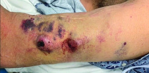
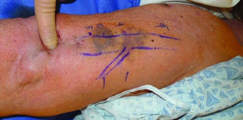
Caption: FIGURE 324-1 Dialysis access health depends on intra-access pressures and needle two recurrences of clinically relevant inflow stenosis in 4 years has low-normal site enlargement. B. In contrast, a right upper arm brachial-cephalic AVF with seven normal to high intra-access pressures with notable needle insertion site enlargement. C. ulcers over 3 years. D. Segmental needle rotation preserves skin integrity even after 7 — FIGURE 324-1 Dialysis access health depends on intra-access pressures and needle insertions. A. A right upper arm brachial-cephalic arteriovenous fistula (AVF) with two recurrences of clinically relevant inflow stenosis in 4 years has low-normal intra-access pressure before and after angioplasty; there is only minimal needle insertion site enlargement. B. In contrast, a right upper arm brachial-cephalic AVF with seven recurrences of cephalic arch outflow stenosis in 4-year cycles between states of high-normal to high intra-access pressures with notable needle insertion site enlargement. C. Focal needle insertions despite available graft segments led to penetrating skin ulcers over 3 years. D. Segmental needle rotation preserves skin integrity even after 7 years of arteriovenous graft (AVG) use.
Figure 2
Caption: FIGURE 324-1 Dialysis access health depends on intra-access pressures and needle two recurrences of clinically relevant inflow stenosis in 4 years has low-normal site enlargement. B. In contrast, a right upper arm brachial-cephalic AVF with seven normal to high intra-access pressures with notable needle insertion site enlargement. C. ulcers over 3 years. D. Segmental needle rotation preserves skin integrity even after 7 — FIGURE 324-2 Anatomy and placement of hemodialysis and peritoneal dialysis catheters. Tunneled catheters have a cuff that creates a barrier between skin flora at the exit site and the sterile catheter tunnel leading into the fibrinous sheath covering the catheter from the vessel insertion point to its tip. Peritoneal dialysis (PD) catheters can be placed fluoroscopically, peritoneoscopically, laparoscopically, and open surgically. The internal cuff is placed within the rectus sheath just laterally to the linea alba, while the external PD catheter cuff should be located 2–4 cm from the skin exit site.
Generated from Harrison's Principles of Internal Medicine, 22nd Edition.