Chapter 184: Leprosy
Infectious Diseases · Part 5 – Infectious Diseases: Bacterial
Detailed clinical reference synthesised from Harrison's Principles of Internal Medicine, 22nd Edition
🔑 Key Clinical Points
- Leprosy (Hansen's disease) is caused by Mycobacterium leprae, an obligate intracellular acid-fast bacterium that infects macrophages and Schwann cells.
- The disease spectrum ranges from tuberculoid (TT) to lepromatous (LL) leprosy based on the host's cell-mediated immune (CMI) response.
- Three cardinal signs for clinical diagnosis: 1) Hypopigmented/erythematous skin lesion with definite loss of sensation, 2) Peripheral nerve thickening with sensory impairment, 3) Positive AFB in slit-skin smear or biopsy.
- WHO disability grading uses the Eye-Hand-Foot (EHF) score: Grade 0 (no impairment), Grade 1 (anesthesia), Grade 2 (visible impairment).
- Incidence dropped significantly after 2000 due to WHO elimination declaration, but transmission continues in India, Brazil, and Indonesia.
- Type 1 reaction (T1R) is a delayed hypersensitivity reaction (reversal reaction) causing acute swelling and nerve damage.
- Type 2 reaction (T2R) is erythema nodosum leprosum (ENL), an immune complex-mediated syndrome causing fever, nodules, and systemic symptoms.
- M. lepromatosis is a distinct species causing diffuse leprosy of Lucio and Latapí, found mainly in Mexico and Central America.
- Slit-skin smear is the standard for classification: Paucibacillary (negative smear, 1-5 lesions) vs Multibacillary (positive smear, 6+ lesions).
- M. leprae cannot be cultured in artificial media; diagnosis relies on slit-skin smear, biopsy, or serology (PGL-1 antibody test).
📑 Table of Contents
- 1. DEFINITION & OVERVIEW
- 1.1 Etiology & Microbiology
- 1.2 M. lepromatosis
- 2. EPIDEMIOLOGY
- 2.1 Transmission & Reservoirs
- 2.2 Incubation & Risk Factors
- 3. ETIOLOGY & PATHOPHYSIOLOGY
- 3.1 Ridley-Jopling Classification
- 4. CLINICAL FEATURES
- 4.1 Clinical Diagnosis
- 4.2 Diagnostic Tools
- 5. DIFFERENTIAL DIAGNOSIS
- 5.1 Diagnostic Clues
- 6. INVESTIGATIONS & DIAGNOSIS
- 6.1 Diagnostic Algorithm
- 7. MANAGEMENT & TREATMENT
- 7.1 Treatment Principles
- 8. PROGNOSIS & COMPLICATIONS
- 8.1 Disability Grading
- 9. SPECIAL CONSIDERATIONS
- 10. KEY PEARLS & CLINICAL TRAPS
- Figures & Illustrations
📋 Figures in This Chapter
| # | Type | Description |
|---|---|---|
| 1 | 🖼 Figure | CHAPTER 184 hands and feet, grade 0 means no anesthesia and no... |
| 2 | 🖼 Figure | Geographic distribution of new leprosy cases, 2022 |
| 3 | 🖼 Figure | Lepromatous (LL) leprosy |
| 4 | 🖼 Figure | Tuberculoid (TT) leprosy |
| 5 | 🖼 Figure | Type 1 leprosy reaction |
| 6 | 🖼 Figure | Tuberculoid (TT) leprosy |
| 7 | 🖼 Figure | Type 1 leprosy reaction |
| 8 | 🖼 Figure | Tuberculoid (TT) leprosy |
1. DEFINITION & OVERVIEW
- Leprosy, also referred to as Hansen's disease, is a chronic infectious disease caused by Mycobacterium leprae.
- Clinical manifestations are largely confined to the skin, peripheral nervous system, eyes, and upper respiratory tract.
- The differing immune responses to M. leprae result in a spectrum of disease ranging from tuberculoid to lepromatous leprosy.
- M. leprae has a predilection for peripheral nerves, and immunologically mediated reactional states can cause nerve damage to the face, arms, and legs.
- This damage often results in disability, which in turn can lead to stigma and social exclusion.
- Harrison's defines this as: 'Leprosy is a neglected disease and is often thought no longer to exist. However, 174,087 new cases from 182 countries were reported in 2022.'
- A general lack of awareness and among both the public and medical practitioners often delays diagnosis and treatment and thus results in irreversible impairments.
- Early diagnosis and treatment of leprosy and leprosy reactions can cure the disease and prevent most chronic complications.
1.1 Etiology & Microbiology
- M. leprae is an obligate, intracellular, acid-fast staining, rod-shaped bacterium.
- Measures 1–8 μm in length and 0.3 μm in diameter.
- M. leprae mostly appears irregularly stained and fragmented or granular, in which case the organism is usually considered to be dead.
- The few bacteria that are brightly and uniformly stained are thought to be solid, viable bacilli.
- The morphologic index is a measure of uniformly stained solid bacilli on slit-skin smear examination and is calculated as the percentage of viable bacilli among the total number of bacilli counted under oil-immersion microscopy.
- The bacteriological index is a logarithmic-scaled measure of the density of bacilli of all forms found in the dermis upon slit-skin smear examination, varying from 0 to 6+ (with or without globi) from the tuberculoid to the lepromatous end of the disease spectrum.
- The bacteriological index falls an average of 1 log unit per year with multidrug therapy.
- M. leprae infects mainly macrophages and Schwann cells.
- It has never been grown in artificial media.
- Reproduction occurs by binary fission, and the organism grows slowly (over 12–14 days) in the footpads of mice.
- The temperature required for survival and proliferation—between 27°C and 30°C—explains the greater impact of the disease on surface areas such as the skin, peripheral nerves, testicles, and upper airways, with less inner visceral involvement.
- M. leprae remains viable for 9 days in the environment.
- Ultrastructural Characteristics: Electron microscopy reveals that M. leprae has a cytoplasm, plasma membrane, cell wall, and capsule.
- The cytoplasm contains structures common in gram-positive microorganisms.
- The plasma membrane has a permeable lipid bilayer containing interacting proteins—the protein surface antigens.
- The cell wall is composed of peptidoglycans bound to branched-chain polysaccharides; these peptidoglycans are arabinogalactans, which support mycolic acids, and lipoarabinomannan (LAM).
- The capsule—the outermost structure—contains lipids, particularly phthiocerol dimycocerosate and phenolic glycolipid (PGL-1), which has a trisaccharide bound to lipid by a molecule of phenol.
- A species-specific phenolic glycolipid, PGL-1, has been identified in M. leprae, and its detection is helpful in serologic diagnosis of leprosy.
- Other varieties of M. leprae antigens identified with monoclonal antibodies include antigens of 18, 28, 7, 14, 36, 65, and 70 kDa that may possibly induce an immune response.
Table 1 — Table 184-1. Ultrastructural Components of M. leprae
| Component | Description | Function/Significance |
|---|---|---|
| Cytoplasm | Contains structures common in gram-positive microorganisms | General bacterial structure |
| Plasma Membrane | Permeable lipid bilayer containing interacting proteins | Contains protein surface antigens |
| Cell Wall | Peptidoglycans bound to branched-chain polysaccharides (arabinogalactans) | Supports mycolic acids and LAM |
| Capsule | Contains lipids, particularly phthiocerol dimycocerosate and phenolic glycolipid (PGL-1) | PGL-1 has a trisaccharide bound to lipid by a molecule of phenol |
1.2 M. lepromatosis
- In 2008, a new mycobacterial species, M. lepromatosis, was isolated from patients with a special type of diffuse lepromatous leprosy known as diffuse leprosy of Lucio and Latapí.
- This clinical variety of leprosy is found mainly in Mexico and Central America.
- M. lepromatosis is very similar to M. leprae microbiologically and clinically.
- Microbiologically, both species are acid-fast and noncultivable and preferentially infect skin and peripheral nerves.
- Clinically, differentiation of M. lepromatosis from M. leprae in individual patients is not diagnostically necessary since both organisms respond well to the same antimycobacterial regimens.
2. EPIDEMIOLOGY
- The true incidence of leprosy is difficult to establish because the figure is very low and the initial signs and symptoms are often insidious, and thus not all cases are detected as they occur.
- In 2022, as stated earlier, the 174,087 new cases were reported to the World Health Organization (WHO) from 182 countries.
- New case detection per year is commonly used as a proxy for incidence, but operational factors, such as the intensity of case detection, the use of surveys, the use of contact tracing, the level of community awareness, and the quality and availability of health care, have a profound effect on case detection rates.
- In nonendemic countries around the world, leprosy is often misdiagnosed simply because it is not considered.
- The registered prevalence of leprosy is defined as the number of patients receiving treatment at a point in time (usually at the end of a calendar year).
- The registered prevalence is a proxy measure for true prevalence, which would include existing cases that have not yet been detected.
- The two factors that determine the registered prevalence are the new case detection rate and the duration of treatment; changes in either factor will affect the registered prevalence.
- The WHO leprosy disability grading system scores patients according to the presence of disabilities of the eyes, hands, and feet.
- For the hands and feet, grade 0 means no anesthesia and no visible impairment; grade 1 signifies anesthesia but no visible impairment; and grade 2 indicates visible impairment.
- For the eyes, grade 0 signifies no eye problems due to leprosy and no evidence of visual loss; grade 1 signifies eye problems due to leprosy without severe effects on vision; and grade 2 indicates severe visual impairment (vision score worse than 6/60; inability to count fingers at 6 meters) and also includes lagophthalmos, iridocyclitis, and corneal opacities.
- The sum score for these six body sites is called the Eye-Hand-Foot (EHF) score and is used as an overall indicator of the impairment status of a person with leprosy.
- Leprosy-related grade 2 disability is usually reported as the proportion of people with such disability at any site among patients newly diagnosed with leprosy in a specific year.
- The global trend in new case detection since 1990 is presented in Fig. 184-1.
- The trend was remarkably static up to the year 2001, with a peak around the year 2000; fell dramatically between 2001 and 2005; and has leveled off from 2006 until 2016.
- Since 2017, a continuous decline has been observed, but the figures for 2020 and 2021 are unreliable due to underreporting during the COVID-19 pandemic.
- The most important factor contributing to the fast downward trend after the year 2000 was the decline in leprosy control activities following the declaration by the WHO in 2000 that leprosy was eliminated as a 'public health problem.'
- Elimination was defined as a prevalence of <1 case per 10,000 population at the global level.
- The decline in new case detection since 2016 to below 200,000 cases per year indicates that transmission of M. leprae is becoming less and that elimination of leprosy could become possible in the next 25 years or so.
- Approximately 40% of all reported leprosy patients are women, but the low proportion in some countries raises concerns about underdiagnosis in women due to poor access to health services, illiteracy, low status, and other cultural factors.
- The age-specific incidence often shows a bimodal pattern, with peaks in the teenage years and in adulthood.
- Around 8% of all newly detected cases are found in children (<15 years of age), a measure that is often taken as an indicator of continued (recent) transmission.
- Leprosy is rare among children <5 years of age.
- Around 5% of all patients have a grade 2 disability.
- There are large variations among world regions and countries in new case detection rates.
- Approximately 80% of global new case detection is reported from India, Brazil, and Indonesia.
- There are also distinct geographic variations within countries, with differences between urban and rural communities and clustering of cases at the village or neighborhood level.
- Geographic variations can be due to differences in health service provision, socioeconomic development, isolation, and poverty.
- Figure 184-2 depicts the geographic distribution of new leprosy cases in 2022.
Table 2 — Table 184-2. WHO Leprosy Disability Grading System (EHF Score)
| Body Site | Grade 0 | Grade 1 | Grade 2 |
|---|---|---|---|
| Hands and Feet | No anesthesia and no visible impairment | Anesthesia but no visible impairment | Visible impairment |
| Eyes | No eye problems due to leprosy and no evidence of visual loss | Eye problems due to leprosy without severe effects on vision | Severe visual impairment (vision score worse than 6/60; inability to count fingers at 6 meters), lagophthalmos, iridocyclitis, and corneal opacities |
2.1 Transmission & Reservoirs
- Understanding of the transmission of M. leprae is limited.
- The existing evidence is largely circumstantial because of the long incubation period from exposure to disease, the inability to culture M. leprae, and the difficulty of diagnosing both infection and early disease.
- M. leprae organisms can be shed in large numbers from the mouth and nose of patients with untreated multibacillary leprosy (droplet infection) and sometimes from damaged skin, but it is unclear whether patients with paucibacillary leprosy can spread the bacillus.
- There is evidence for transmission between humans and—in southern U.S. states—for zoonotic transmission through wild armadillos.
- The main route of entry into the body is assumed to be the respiratory tract, but in patients with wounds or tattoos, transmission through the skin also is possible.
- It is assumed that humans are the main reservoir of infection for M. leprae.
- The armadillo is also a reservoir for human infection.
- Certain species of monkeys and red squirrels are infected with M. leprae in the wild, but there is no evidence of transmission to humans through contact with these animals.
- Evidence is weak for the potential of water and soil as environmental sources of M. leprae.
- The higher incidence rate of leprosy among household contacts of multibacillary cases than among those of paucibacillary cases suggests that multibacillary cases represent an important reservoir for undetected and untreated cases in the community.
- That is, a prolonged period between the onset of signs of leprosy and treatment due to a delay in diagnosis and initiation of multidrug therapy increases exposure in the community.
- Persons with subclinical leprosy are likely to be a main source of infection, given that multidrug therapy for clinical leprosy apparently has not made an impact on transmission.
2.2 Incubation & Risk Factors
- The incubation period of leprosy is estimated to range from 2 to ≥10 years.
- The incubation period for multibacillary leprosy appears to be longer (5 to ≥15 years) than that for paucibacillary leprosy (~2–5 years).
- Poverty-associated factors such as low level of education, poor hygiene, and food shortages have been identified as risk factors for leprosy.
- But the most important risk factors are associated with intimacy and duration of contact with a leprosy patient, in particular with an index case with multibacillary leprosy, and the intensity of contact with and physical distance from the index patient.
- Increasing evidence from studies in twins and from observational studies supports host genetic susceptibility to leprosy.
- Ongoing studies are exploring the mechanism underlying genetic susceptibility to leprosy and its clinical manifestations.
3. ETIOLOGY & PATHOPHYSIOLOGY
- Whatever the route of M. leprae's entry into the human body, the pathogenic process usually starts in the peripheral nerves.
- Once bacilli are engulfed by Schwann cells, the histopathologic changes in nerve and skin—and thus the type of leprosy that develops—depend on the immunologic resistance of the person infected, in particular on the cell-mediated immune (CMI) response to the bacillus and its antigens.
- Immunologic resistance is strong at the tuberculoid end of the spectrum, gradually diminishes through the borderline spectrum, and is weakest in lepromatous leprosy.
- The LL and TT types of leprosy are relatively stable, with little or no change in clinical disease expression over time, while the BL, BB, and BT types are unstable both clinically and immunologically.
- Further distinction indicates that subpolar types of TT and LL leprosy (TTs and LLs) are less stable than polar types (TTp and LLp).
- The immune reaction depends on predisposing genetic factors and the extent of exposure to M. leprae.
- The host tissue's reaction and related damage are largely due to delayed hypersensitivity.
- In response to the presence of M. leprae, a granuloma is formed either by macrophage–lymphocyte interaction when there is immunity or otherwise by macrophages only.
- The formation of a granuloma is preceded by a stage of infiltration by lymphocytes alone, as is seen in IL.
- Because of the strong immune response toward the tuberculoid end of the spectrum, macrophages, along with many lymphocytes, become fixed epithelioid cells, and groups of these cells become giant cells.
- The tuberculoid granuloma leads to nerve destruction resulting in anesthesia and muscle weakness.
- The cellular response is less focal and less destructive in the borderline portion of the spectrum; consequently, there is less damage to nerves and few bacilli are present.
- In BL leprosy, there are macrophage granulomas along with lymphocytes, but little to nerve damage and more bacilli.
- In LL leprosy, bacilli multiply within macrophages and outside macrophages either singly or in small groups.
- The bacilli are packed within macrophages called globi and outside macrophages either singly or in small groups.
- Inside these cells, M. leprae multiplies in unrestricted fashion, and hundreds of bacilli may be present; the organisms are arranged in parallel arrays placed side by side as a result of the presence of surface lipids (glial substances).
- Liberated bacilli from these cells are engulfed by histiocytes, becoming wandering macrophages and traveling throughout the body to other nerves and tissues via blood, lymph, and tissue fluids.
- In addition, there are diffuse lepromas in LL leprosy that consist of histiocytes and/or macrophages, with very few lymphocytes and plasma cells.
3.1 Ridley-Jopling Classification
- In 1962, Ridley and Jopling described five overlapping categories of leprosy: tuberculoid (TT), borderline tuberculoid (BT), mid-borderline (BB), borderline lepromatous (BL), and lepromatous (LL).
- An early clinical manifestation is recognized and referred to as indeterminate leprosy (IL).
- The WHO has therefore introduced a simplified classification system based on slit-skin smear: patients with negative slit-skin smear results at all body sites are classified as having paucibacillary leprosy, whereas patients with positive smears at any body site are classified as having multibacillary leprosy.
- However, because slit-skin smear facilities are not available or dependable in many countries, most leprosy control programs use clinical criteria only for classifying leprosy and deciding on the appropriate treatment regimen for individual patients.
- In this circumstance, paucibacillary leprosy is defined as one to five skin lesions and no or only one involved peripheral nerve, while multibacillary leprosy is defined as six or more skin lesions and/or more than one involved peripheral nerve.
Table 3 — Table 184-3. Ridley-Jopling Classification of Leprosy
| Type | Immunologic Status | Stability | Clinical Features |
|---|---|---|---|
| Tuberculoid (TT) | Strong CMI | Stable | Single or few lesions (up to three), well-defined edge, complete loss of sensation, thickened nerves |
| Borderline Tuberculoid (BT) | Moderate CMI | Unstable | 3-9 lesions, asymmetric, variable margins, less intense sensation loss, susceptible to T1R |
| Mid-Borderline (BB) | Intermediate CMI | Unstable | Multiple plaque lesions, bilateral, symmetrical, moderate sensation loss, variable nerve thickening |
| Borderline Lepromatous (BL) | Weak CMI | Unstable | Numerous bilateral lesions, 2-3 cm diameter, coppery hue, minimal sensation loss, few bacilli |
| Lepromatous (LL) | Weak/No CMI | Stable (Polar) / Unstable (Subpolar) | Innumerable bilateral lesions, diffuse induration, shiny surface, no sensation loss, high bacillary load |
4. CLINICAL FEATURES
- Leprosy is a disease affecting mainly the skin, cutaneous and peripheral nerves, mucous membranes, and, less commonly, other sites such as joints, lymph nodes, eyes, and testes.
- Other systemic manifestations may occur, particularly in BL and LL disease, with or without leprosy reactions.
- Most dermal and cutaneous nerves feeding skin lesions are affected—e.g., the supraorbital, great auricular, radial cutaneous, infrapatellar, superficial fibular, and sural nerves and the cutaneous nerves of the thigh.
- The peripheral nerves involved include the ulnar, median, radial (in upper limbs), lateral popliteal, and posterior tibial (in lower limbs).
- The cranial nerves commonly involved are the trigeminal and facial.
- Indeterminate Leprosy (IL): This early clinical type manifests as one or a few hypopigmented or faintly erythematous, ill-defined or well-defined macular lesions measuring 1–5 cm in diameter.
- These lesions invariably occur on the external aspects of the limbs, buttocks, and face, with mild to moderate impairment of touch and/or thermal sensations.
- There is no thickening of the corresponding cutaneous and peripheral nerves.
- IL is often, but not always, the first clinical sign of leprosy.
- This type either heals spontaneously or progresses to a determinate form of the disease (TT, BT, BB, BL, or LL), depending on CMI status.
- Tuberculoid (TT) Leprosy: TT leprosy presents lesions either as a well-defined, hypopigmented macule or as a raised, erythematous/brown/copper-colored plaque with a well-defined edge.
- The lesions may be found on any part of the skin and are characterized by complete loss of fine touch and temperature sensations over their surface.
- Skin lesions are single or few (up to three) in number and can be of any size, but they seldom measure >10 cm in diameter.
- In plaque-type lesions, the raised clear-cut edge often slopes inward to a flattened and sometimes hypopigmented central area, acquiring an annular configuration.
- The skin surface of both macular and plaque lesions is dry, hairless, and anesthetic because of destruction of underlying superficial cutaneous nerves.
- Larger corresponding cutaneous nerves are thickened in a limited number of cases.
- On the face, sensory impairment may be difficult to demonstrate because of the generous and bilateral supply of sensory nerve endings.
- Autonomic nerve damage within the lesion is responsible for surface dryness and loss of sweating over the lesion.
- A solitary peripheral-nerve trunk in the vicinity of a lesion may be thickened, with sensory loss of the area supplied and with or without motor disfigurement.
- Borderline Tuberculoid (BT) Leprosy: BT leprosy is characterized by either macular or plaque-type lesions numbering three to nine or more and asymmetrically located on any part of the body, with variable sizes and contours.
- The margins of the lesions range from poorly defined to well defined; sometimes both forms of margin are seen in one lesion.
- There may be smaller satellite lesions around a larger one, especially on sides where the margin is less defined; this characteristic indicates downgrading of the lesion from TT to BT leprosy.
- The edges of plaque lesions may slope outward in contrast to TT lesions, which slope inward; plaques may gradually fade outward and eventually blend into normal-looking skin.
- Loss of sensation is less intense than it is in TT lesions and dryness on the surface less conspicuous.
- Several peripheral nerves are likely to be enlarged in an asymmetrical pattern, with sensory and motor deficits.
- One of the most striking features of BT leprosy is susceptibility to a type 1 leprosy reaction (T1R; see below) that exacerbates skin lesions and/or peripheral nerves.
- If not diagnosed and treated early, disease in these patients tends to downgrade across the spectrum to BB, BL, or LLs leprosy, with an increasing bacteriologic index and a regressed CMI response causing nerve damage along the way.
- Mid-Borderline (BB) Leprosy: This form of leprosy is unstable.
- Many cases downgrade toward BL and LL disease, especially if not treated.
- There are multiple plaque lesions and, not infrequently, macular lesions; the lesions are of various shapes and sizes, are bilateral, and usually occur in a more or less symmetrical distribution.
- In annular lesions, the inner edge is well demarcated and 'punched out,' and the outer edge is ill defined and merges with normal-looking skin.
- The surface of the lesions is moderately shiny, and the central area looks pale.
- There is minimal loss of sensation over the lesions.
- Nerve damage is variable in BB leprosy.
- Many nerves may be thickened, and this effect may be asymmetrical.
- BB leprosy is not commonly observed and rapidly changes its spectrum—rarely to BT leprosy but more often to BL disease.
- The lepromin test is negative.
- Slit-skin smears of lesions show a moderate number of AFB (2+ to 3+).
- Borderline Lepromatous (BL) Leprosy: In BL leprosy, there are numerous bilateral, round or oval, macular, diffusely infiltrated, erythematous or hypopigmented lesions with moderately defined borders.
- The lesions are usually 2–3 cm in diameter, may have a coppery hue, and tend to become symmetrical.
- Some loss of lesion is responsible for surface dryness and loss of sweating over the lesion.
- A solitary peripheral-nerve trunk in the vicinity of a lesion may be thickened, with sensory loss of the area supplied and with or without motor disfigurement.
- On slit-skin smear examination, no acid-fast bacilli (AFB) are normally found.
- The lepromin skin test is strongly positive, signifying good host CMI status.
- Lepromatous (LL) Leprosy: LL leprosy presents with innumerable bilateral, symmetrically distributed, diffusely indurated, erythematous, copper-colored or skin-colored patches or plaques.
- There is no loss of sensation over these lesions, which have a smooth, shiny surface.
- The lesions spread over the face, earlobes, ears, extensor aspects of the upper and lower extremities, back, and buttocks.
- Induration can readily be recognized when lesions are viewed tangentially under natural sunlight.
- The induration initially is of a finer type but gradually becomes coarse, and lesions then progress to papules, plaques, and nodules.
- Bilateral earlobe thickening and eyebrow loss occur.
- Coarse induration on the face sometimes results in gross skin folds that lead to an appearance referred to as 'lion face,' particularly when associated with loss of eyebrows and thickening of earlobes.
- Of all cases of LL leprosy, 10–15% are of the polar type (LLp) from the time of lesion onset; the remaining cases downgrade from the untreated borderline spectrum to subpolar LLs leprosy.
- Patients with LLs disease develop nerve damage during the borderline stages.
- In LLp disease, involvement of peripheral nerves occurs late and is bilateral and symmetrical, with sensory loss in a 'glove-and-stocking' distribution.
- Slit-skin smear examination shows a bacteriologic index of 4+ to 6+ with globi.
- Primary Neuritic Leprosy: In some countries, such as India and Nepal, primary neuritic disease is observed in 2–10% of all leprosy cases, with only peripheral nerve involvement and no skin lesions.
- Nerve thickening and sensory loss occur in the affected area, with or without a motor deficit.
- With disease progression, papules, nodules, and plaques develop over the macular lesions.
- In untreated patients, new ill-defined skin lesions continue to develop.
- Widespread but asymmetrical thickening of peripheral nerves, with or without tenderness, leads to sensory and motor deficits.
- The lepromin test gives negative results, as it does in all degrees of lepromatous leprosy.
- Slit-skin smear examination of lesions shows a bacteriologic index varying from 3+ to 4+.
- Histoid Leprosy: Histoid leprosy is a rare form of LL leprosy in which waxy, shiny, firm, symmetrical or asymmetrical nodules and plaques are observed over normal-looking skin.
- Histologic examination of these lesions shows specific spindle-cell granulomas.
- Slit-skin smear examination reveals high bacteriologic and microbiologic indices without globi in most cases.
- Diffuse Leprosy of Lucio and Latapí: This rare form of nonnodular LL leprosy occurring in Mexico and Central America is characterized by diffuse shiny infiltration of the skin and widespread sensory loss.
- The skin looks waxy and has a shiny appearance ('lepra bonita,' or beautiful leprosy), with obvious diffuse induration of the earlobes and forehead as well as loss of eyebrows, sometimes eyelashes, and not infrequently all body hair.
- This form of leprosy can be complicated by an unusual reaction known as Lucio's phenomenon (see below).
- Systemic Involvement: In LL leprosy, AFB are found in the lymph nodes, spleen, liver, bone marrow, adrenal glands, smooth and striated muscles, tooth pulp, testes, oral cavity, nose, larynx, and eyes.
- Involvement of the testes leads first to sterility and then to gynecomastia and impotence.
- Eye involvement includes corneal anesthesia; early on, this manifestation is due to bacillary infiltration of corneal nerves, while later it arises from damage to the ophthalmic division of the trigeminal nerve.
- In addition, eye involvement includes episcleritis, iridocyclitis, iris atrophy, cataract and glaucoma, lagophthalmos, corneal ulceration and perforation, and blindness.
- The nose is a portal of entry for M. leprae and is the earliest site of involvement in LL leprosy.
- Edema and mucosal thickening occur in the inferior turbinate and nasal septum, with crusting and epistaxis.
- Later, patients develop chronic rhinitis with loss of smell sensation.
- Septal perforation due to bony destruction, with typical saddle-nose disfigurement, is common in advanced LL disease.
- In late-stage LL leprosy, ulceration of the tongue, pharynx, hard and soft palates (leading to palate perforation), tonsillar pillars, and uvula occurs.
4.1 Clinical Diagnosis
- Three cardinal signs indicate a diagnosis of leprosy.
- The diagnosis can be established when at least one of these three signs is present:
-
- Hypopigmented or erythematous skin lesion(s) with definite loss or impairment of sensation: The clinical presentation of skin patches or plaques is diagnostic when it is associated with a definite loss or impairment of sensation (light touch, pain, and/or temperature).
- Diagnostic dilemmas arise in the indeterminate stage of leprosy because of variable loss of sensation and the presence of facial lesions (i.e., because the density of innervation in the face can compensate for damage to certain nerve branches).
-
- Involvement of the peripheral nerves, as demonstrated by definite thickening with sensory impairment: Thickening of a peripheral nerve should be assessed by palpation of the affected nerve and a comparison with the corresponding contralateral nerve.
- In multibacillary leprosy, thickening of nerves is often bilateral.
- Nerve tenderness is established by the application of mild pressure on the nerve during palpation with the fingertips.
- The peripheral nerves commonly palpated in a leprosy patient are the greater auricular, ulnar, radial, radial cutaneous, median, lateral popliteal, posterior tibial, sural, and superficial peroneal nerves.
-
- A positive result for AFB in slit-skin smears, establishment of the presence of AFB in a skin smear or biopsy sample, or a positive result in a biopsy polymerase chain reaction (PCR).
- Testing of Skin Sensation: Light-touch sensation is tested with cotton, wool, or a feather.
- Pain is assessed as the patient's ability to distinguish between the sharp and blunt ends of a wooden or bamboo toothpick.
- Thermal sensation thresholds are assessed with computer-assisted sensory testing equipment.
4.2 Diagnostic Tools
- SLIT-SKIN SMEAR: Normally a slit-skin smear is taken from four sites: the right earlobe, the forehead above the eyebrows, the chin, and the left buttock in men or the left upper thigh in women.
- The material is stained with Ziehl-Neelsen reagent and examined with a light microscope.
- The bacteriological index is determined with a standard logarithmic scale and graded from 0 to 6.
- The microbiologic index is determined as the percentage of solid, stained AFB.
- SKIN BIOPSY: A skin biopsy is done to confirm the diagnosis of leprosy, to classify the disease, to support the diagnosis of reactions, and to determine cure after the completion of multidrug therapy.
- When macular lesions are suspected of reflecting IL, a biopsy sample should be taken from the middle of a lesion; with plaques, a sample should be obtained from the active indurated edge.
- When there are numerous skin lesions with different morphologies, more than one biopsy sample is required for proper evaluation of the disease spectrum.
- Identification of early lesions by histopathologic techniques is enhanced by immunochromatographic staining, which reveals the presence of M. leprae antigens.
- PGL-1 ANTIBODY TEST: PGL-1 is a specific lipid on the M. leprae cell wall.
- A PGL-1 enzyme-linked immunosorbent assay (ELISA) has been used for serologic diagnosis of leprosy, yielding positive results in 90–95% of multibacillary cases and in 25–60% of paucibacillary cases.
5. DIFFERENTIAL DIAGNOSIS
- Differential dilemmas arise in the indeterminate stage of leprosy because of variable loss of sensation and the presence of facial lesions.
- The density of innervation in the face can compensate for damage to certain nerve branches.
- Clinical diagnosis relies on the three cardinal signs: skin lesion with sensory loss, nerve thickening with sensory impairment, and positive AFB in slit-skin smear or biopsy.
- Indeterminate leprosy lesions may mimic other dermatological conditions such as vitiligo, tinea versicolor, or post-inflammatory hypopigmentation, but the key differentiating feature is the loss of sensation.
- Nerve thickening is a hallmark of leprosy and distinguishes it from other causes of skin hypopigmentation.
- Positive AFB in slit-skin smear or biopsy confirms the diagnosis and distinguishes it from non-infectious causes of nerve damage.
5.1 Diagnostic Clues
- Pathognomonic signs: Hypopigmented or erythematous skin lesion(s) with definite loss or impairment of sensation.
- Hallmark finding: Thickening of a peripheral nerve with sensory impairment.
- Key feature: Positive result for AFB in slit-skin smears, establishment of the presence of AFB in a skin smear or biopsy sample, or a positive result in a biopsy polymerase chain reaction (PCR).
- Clinical clues: Lesions invariably occur on the external aspects of the limbs, buttocks, and face.
- Red flags: 'Silent neuritis' or 'quiet nerve paralysis' is defined as progressive sensory or motor impairment in the absence of symptoms such as pain, paresthesia, or tenderness of the nerve and with no obvious signs of leprosy reactions.
- Eponymous signs: 'Lion face' appearance associated with loss of eyebrows and thickening of earlobes in LL leprosy.
- Clinical pearls: The bacteriological index falls an average of 1 log unit per year with multidrug therapy.
6. INVESTIGATIONS & DIAGNOSIS
- Clinical Diagnosis: Three cardinal signs indicate a diagnosis of leprosy.
- The diagnosis can be established when at least one of these three signs is present.
-
- Hypopigmented or erythematous skin lesion(s) with definite loss or impairment of sensation: The clinical presentation of skin patches or plaques is diagnostic when it is associated with a definite loss or impairment of sensation (light touch, pain, and/or temperature).
- Diagnostic dilemmas arise in the indeterminate stage of leprosy because of variable loss of sensation and the presence of facial lesions (i.e., because the density of innervation in the face can compensate for damage to certain nerve branches).
-
- Involvement of the peripheral nerves, as demonstrated by definite thickening with sensory impairment: Thickening of a peripheral nerve should be assessed by palpation of the affected nerve and a comparison with the corresponding contralateral nerve.
- In multibacillary leprosy, thickening of nerves is often bilateral.
- Nerve tenderness is established by the application of mild pressure on the nerve during palpation with the fingertips.
- The peripheral nerves commonly palpated in a leprosy patient are the greater auricular, ulnar, radial, radial cutaneous, median, lateral popliteal, posterior tibial, sural, and superficial peroneal nerves.
-
- A positive result for AFB in slit-skin smears, establishment of the presence of AFB in a skin smear or biopsy sample, or a positive result in a biopsy polymerase chain reaction (PCR).
- Diagnostic Tools: TESTING OF SKIN SENSATION: Light-touch sensation is tested with cotton, wool, or a feather.
- Pain is assessed as the patient's ability to distinguish between the sharp and blunt ends of a wooden or bamboo toothpick.
- Thermal sensation thresholds are assessed with computer-assisted sensory testing equipment.
- SLIT-SKIN SMEAR: Normally a slit-skin smear is taken from four sites: the right earlobe, the forehead above the eyebrows, the chin, and the left buttock in men or the left upper thigh in women.
- The material is stained with Ziehl-Neelsen reagent and examined with a light microscope.
- The bacteriological index is determined with a standard logarithmic scale and graded from 0 to 6.
- The microbiologic index is determined as the percentage of solid, stained AFB.
- SKIN BIOPSY: A skin biopsy is done to confirm the diagnosis of leprosy, to classify the disease, to support the diagnosis of reactions, and to determine cure after the completion of multidrug therapy.
- When macular lesions are suspected of reflecting IL, a biopsy sample should be taken from the middle of a lesion; with plaques, a sample should be obtained from the active indurated edge.
- When there are numerous skin lesions with different morphologies, more than one biopsy sample is required for proper evaluation of the disease spectrum.
- Identification of early lesions by histopathologic techniques is enhanced by immunochromatographic staining, which reveals the presence of M. leprae antigens.
- PGL-1 ANTIBODY TEST: PGL-1 is a specific lipid on the M. leprae cell wall.
- A PGL-1 enzyme-linked immunosorbent assay (ELISA) has been used for serologic diagnosis of leprosy, yielding positive results in 90–95% of multibacillary cases and in 25–60% of paucibacillary cases.
- Because filaments are not available in most peripheral health centers, the WHO recommends that a ballpoint pen be used instead.
- The testing protocol is the same as in the SWM test: the stimulus is delivered by touching the test sites with the tip of a ballpoint pen held at an angle.
6.1 Diagnostic Algorithm
- Step 1: Clinical Assessment
- Look for hypopigmented or erythematous skin lesion(s) with definite loss or impairment of sensation.
- Assess peripheral nerve thickening with sensory impairment.
- Check for positive AFB in slit-skin smears or biopsy.
- Step 2: Classification
- If slit-skin smear is negative at all sites: Classify as Paucibacillary (PB).
- If slit-skin smear is positive at any site: Classify as Multibacillary (MB).
- If clinical criteria only: PB = 1-5 lesions and no or only one involved peripheral nerve; MB = 6+ lesions and/or more than one involved peripheral nerve.
- Step 3: Confirmatory Testing
- Perform slit-skin smear from four sites (right earlobe, forehead, chin, left buttock/thigh).
- Perform skin biopsy if diagnosis is uncertain or for reaction classification.
- Perform PGL-1 ELISA if available.
- Step 4: Reaction Assessment
- Evaluate for Type 1 (T1R) or Type 2 (T2R) reactions.
- Assess nerve function impairment and disability (EHF score).
7. MANAGEMENT & TREATMENT
- Early diagnosis and treatment of leprosy and leprosy reactions can cure the disease and prevent most chronic complications.
- Multidrug therapy (MDT) is the standard of care.
- The bacteriological index falls an average of 1 log unit per year with multidrug therapy.
- Leprosy reactions are immunologic phenomena that occur before, during, or after treatment.
- They are severe complications that need to be diagnosed and treated early to prevent nerve function impairment and subsequent disfigurement as well as blindness.
- Type 1 Leprosy Reaction (T1R): T1R is a delayed hypersensitivity reaction associated with sudden alteration of CMI status and leading to a shift in the patient's position on the leprosy spectrum.
- T1R is also called a reversal reaction because of the upgrading of CMI status.
- T1R is usually observed in the borderline portion of the spectrum.
- Skin lesions are characterized by acute swelling and redness.
- Nerves may be painful and tender because of neuritis, with consequent nerve damage and disfigurement.
- Loss of nerve function can be much less obvious than usual when it occurs without other signs of inflammation.
- This 'silent neuritis' may lead to sensory and motor impairment in the hands, feet, and face.
- Arthralgia or arthritis sometimes occurs.
- Rarely, the patient may develop fever and malaise, tenosynovitis, and edema of the feet and hands.
- In the severe form of T1R, nerve abscesses may be formed.
- Type 2 Leprosy Reaction (T2R): T2R, also known as ENL (erythema nodosum leprosum), is an immune complex–mediated syndrome (i.e., an antigen–antibody reaction involving complement) and leaves an ulcer of irregular shape may follow later.
- The ulcer heals, leaving a superficial scar.
- Patients remain afebrile throughout.
- This reaction occurs mostly during multidrug therapy but can also develop in untreated patients.
- Evanescent, pink-to-red, maculopapular, papular, nodular, or plaque lesions suddenly appear and are usually accompanied by constitutional symptoms like malaise and fever, with or without painful swelling in the joints.
- These crops of skin lesions present on the outer aspects of the thighs, legs, and face.
- They are painful or tender and warm, blanch with light finger pressure, and last for a few days.
- The lesions change in color from pink/red to bluish and brownish after 24–48 h and turn dark in a week.
- Rarely, ENL lesions become vesicular, pustular, bullous, and necrotic and break down to produce ulceration (erythema nodosum necroticans).
- The patient may have other associated signs such as lymph node enlargement, myositis, arthritis, synovitis, rhinitis, epistaxis, laryngitis, iridocyclitis, glaucoma, painful dactylitis, acute epididymoorchitis, nephritis and renal failure, hepatosplenomegaly, anemia, and—at a later stage—amyloidosis.
- Severe T2R may include swollen, painful, and tender nerve trunks with sensory and motor deficits.
- Lucio's Phenomenon: Lucio's phenomenon is observed in diffuse leprosy of Lucio and Latapí and may be a variant of erythema nodosum necroticans.
- Marked vasculitis and thrombosis of the superficial and deep vessels result in hemorrhage and infarction of the skin.
- Clinically, the skin reaction begins as slightly indurated, bluish-red, ill-defined, painful, and rarely palpable plaques with an erythematous halo, usually developing on one limb but sometimes on other areas of the body.
- The lesions are irregular or triangular.
- After a few days, they become purplish at the center; a central hemorrhagic infarct may develop with or without blister formation, and a necrotic eschar that detaches easily may follow later.
- Nerve Function Impairment, Neuritis, and Disfigurement: The terms nerve function impairment, nerve damage, neuropathy, and neuritis are often used interchangeably for the sensory, motor, and/or autonomic nerve deficits that occur because of the pathologic processes resulting from M. leprae infection of the nerve.
- Neuritis (nerve inflammation) in leprosy is usually a subacute, demyelinating, and unremitting event involving cutaneous nerves and larger peripheral nerves.
- 'Silent neuritis' or 'quiet nerve paralysis' is defined as progressive sensory or motor impairment in the absence of symptoms such as pain, paresthesia, or tenderness of the nerve and with no obvious signs of leprosy reactions.
- Neuritis can occur at any time during leprosy but is more common and severe during leprosy reactions, mainly in T1R.
- Sensory and motor neuropathy can lead to secondary impairments in the upper and lower extremities, such as muscle atrophy, mobile- and fixed-joint contractures, bone absorption of digits, and cracks and wounds.
- Because filaments are not available in most peripheral health centers, the WHO recommends that a ballpoint pen be used instead.
- The testing protocol is the same as in the SWM test: the stimulus is delivered by touching the test sites with the tip of a ballpoint pen held at an angle.
7.1 Treatment Principles
- Multidrug therapy (MDT) is the standard of care.
- The bacteriological index falls an average of 1 log unit per year with multidrug therapy.
- Early diagnosis and treatment of leprosy and leprosy reactions can cure the disease and prevent most chronic complications.
- Leprosy reactions are immunologic phenomena that occur before, during, or after treatment.
- They are severe complications that need to be diagnosed and treated early to prevent nerve function impairment and subsequent disfigurement as well as blindness.
- Glucocorticoid treatment should be given when nerve function impairment is diagnosed.
8. PROGNOSIS & COMPLICATIONS
- The decline in new case detection since 2016 to below 200,000 cases per year indicates that transmission of M. leprae is becoming less and that elimination of leprosy could become possible in the next 25 years or so.
- Around 5% of all patients have a grade 2 disability.
- Sensory and motor neuropathy can lead to secondary impairments in the upper and lower extremities, such as muscle atrophy, mobile- and fixed-joint contractures, bone absorption of digits, and cracks and wounds.
- Leprosy reactions are severe complications that need to be diagnosed and treated early to prevent nerve function impairment and subsequent disfigurement as well as blindness.
- Severe T2R may include swollen, painful, and tender nerve trunks with sensory and motor deficits.
- In the severe form of T1R, nerve abscesses may be formed.
- Lucio's phenomenon results in hemorrhage and infarction of the skin.
- Systemic involvement in LL leprosy can lead to sterility, gynecomastia, impotence, and blindness.
8.1 Disability Grading
- The WHO leprosy disability grading system scores patients according to the presence of disabilities of the eyes, hands, and feet.
- For the hands and feet, grade 0 means no anesthesia and no visible impairment; grade 1 signifies anesthesia but no visible impairment; and grade 2 indicates visible impairment.
- For the eyes, grade 0 signifies no eye problems due to leprosy and no evidence of visual loss; grade 1 signifies eye problems due to leprosy without severe effects on vision; and grade 2 indicates severe visual impairment (vision score worse than 6/60; inability to count fingers at 6 meters) and also includes lagophthalmos, iridocyclitis, and corneal opacities.
- The sum score for these six body sites is called the Eye-Hand-Foot (EHF) score and is used as an overall indicator of the impairment status of a person with leprosy.
- Leprosy-related grade 2 disability is usually reported as the proportion of people with such disability at any site among patients newly diagnosed with leprosy in a specific year.
9. SPECIAL CONSIDERATIONS
- The text does not explicitly detail special considerations for pregnancy, pediatrics, elderly, renal/hepatic impairment, or immunocompromised states in the provided source text.
- However, the text notes that around 8% of all newly detected cases are found in children (<15 years of age), a measure that is often taken as an indicator of continued (recent) transmission.
- Leprosy is rare among children <5 years of age.
- The text mentions that in nonendemic countries around the world, leprosy is often misdiagnosed simply because it is not considered.
10. KEY PEARLS & CLINICAL TRAPS
- Board Exam Favorite: The three cardinal signs for clinical diagnosis of leprosy are: 1) Hypopigmented/erythematous skin lesion with definite loss of sensation, 2) Peripheral nerve thickening with sensory impairment, 3) Positive AFB in slit-skin smear or biopsy.
- Clinical Trap: Indeterminate leprosy lesions may mimic vitiligo or tinea versicolor; the key differentiating feature is the loss of sensation.
- Clinical Trap: 'Silent neuritis' or 'quiet nerve paralysis' is defined as progressive sensory or motor impairment in the absence of symptoms such as pain, paresthesia, or tenderness of the nerve and with no obvious signs of leprosy reactions.
- Mnemonic: 'Leprosy Loves Nerves' (Skin, Peripheral Nerves, Eyes, Upper Respiratory Tract).
- Clinical Pearl: The bacteriological index falls an average of 1 log unit per year with multidrug therapy.
- Clinical Pearl: Of all cases of LL leprosy, 10–15% are of the polar type (LLp) from the time of lesion onset; the remaining cases downgrade from the untreated borderline spectrum to subpolar LLs leprosy.
- Clinical Pearl: The WHO leprosy disability grading system uses the Eye-Hand-Foot (EHF) score.
- Clinical Pearl: M. lepromatosis is a distinct species causing diffuse leprosy of Lucio and Latapí, found mainly in Mexico and Central America.
Figures & Illustrations
Reproduced from Harrison's 22nd Edition.
Figure 1

Caption: CHAPTER 184 hands and feet, grade 0 means no anesthesia and no visible impair- and poverty. Figure 184-2 depicts the geographic distribution of new ment; grade 1 signifies anesthesia but no visible impairment; and grade leprosy cases in 2022. 2 indicates visible impairment. For the eyes, grade 0 signifies no eye problems due to leprosy and no evidence of visual loss; grade 1 signifies Transmission Understanding of the transmission of M. leprae eye problems due to leprosy without severe effects on vision; and grade is limited. The existing evidence is largely circumstantial because of 2 indicates severe visual impairment (vision score worse than 6/60; the long incubation period from exposure to disease, the inability to inability to count fingers at 6 meters) and also includes lagophthalmos, culture M. leprae, and the difficulty of diagnosing both infection and iridocyclitis, and corneal opacities. The sum score for these six body early disease. M. leprae organisms can be shed in large numbers from sites is called the Eye-Hand-Foot (EHF) score and is used as an overall the mouth and nose of patients with untreated multibacillary leprosy — Figure 184-1: Global trend in leprosy new-case detection from 1990 to 2022, showing a peak around 2000, a dramatic decline between 2001-2005, and a continuous decline since 2017.
Figure 2

Caption: FIGURE 184-2 Geographic distribution of new leprosy cases, 2022. (Reproduced with to elimination. Wkly Epidemiol Rec 98:409, 2023.) — Figure 184-2: Geographic distribution of new leprosy cases in 2022, highlighting high-burden regions in India, Brazil, and Indonesia.
Figure 3

Caption: FIGURE 184-6 Lepromatous (LL) leprosy. Multiple nodules on ears and face and loss of eyebrows. (From Dr. K. Mponda, Department of Dermatology, Queen Elisabeth Central Hospital, Blantyre, Malawi, with permission.) — Figure 184-3: Tuberculoid (TT) leprosy presenting as a hypopigmented macular lesion with a well-defined edge and loss of fine-touch sensation.
Figure 4
Caption: FIGURE 184-3 Tuberculoid (TT) leprosy. Hypopigmented macular lesion with a well- FIGURE 184-4 Borderline tuberculoid (BT) leprosy. Macular lesion with irregular, defined edge and loss of fine-touch sensation. (From Dr. H. K. Kar, with permission.) moderately defined edge and satellite lesion, with loss of sensation. (From Dr. W. H. van Brakel, with permission from NLR.) and face, with mild to moderate impairment of touch and/or thermal sensations. There is no thickening of the corresponding cutaneous Mid-Borderline (BB) Leprosy This form of leprosy is unstable. and peripheral nerves. IL is often, but not always, the first clinical Many cases downgrade toward BL and LL disease, especially if not sign of leprosy. This type either heals spontaneously or progresses to treated. There are multiple plaque lesions and, not infrequently, macu- a determinate form of the disease (TT, BT, BB, BL, or LL), depending lar lesions; the lesions are of various shapes and sizes, are bilateral, and on CMI status. usually occur in a more or less symmetrical distribution. In annular — Figure 184-4: Borderline tuberculoid (BT) leprosy showing a macular lesion with irregular, moderately defined edge and satellite lesions, with loss of sensation.
Figure 5

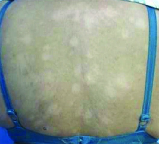
Caption: FIGURE 184-7 Type 1 leprosy reaction. Increased inflammation of existing lesions. (From Dr. W. H. van Brakel, with permission from NLR.) This reaction occurs mostly during multidrug therapy but can also develop in untreated patients. Evanescent, pink-to-red, maculopapular, papular, nodular, or plaque lesions suddenly appear and are usually accompanied by constitutional symptoms like malaise and fever, with 1. or without painful swelling in the joints (Fig. 184-8). These crops of skin lesions present on the outer aspects of the thighs, legs, and face. They are painful or tender and warm, blanch with light finger pres- — Figure 184-5: Borderline lepromatous (BL) leprosy characterized by numerous diffusely infiltrated erythematous and hypopigmented macules, downgrading from borderline tuberculoid to lepromatous leprosy.
Figure 6

Caption: FIGURE 184-3 Tuberculoid (TT) leprosy. Hypopigmented macular lesion with a well- FIGURE 184-4 Borderline tuberculoid (BT) leprosy. Macular lesion with irregular, defined edge and loss of fine-touch sensation. (From Dr. H. K. Kar, with permission.) moderately defined edge and satellite lesion, with loss of sensation. (From Dr. W. H. van Brakel, with permission from NLR.) and face, with mild to moderate impairment of touch and/or thermal sensations. There is no thickening of the corresponding cutaneous Mid-Borderline (BB) Leprosy This form of leprosy is unstable. and peripheral nerves. IL is often, but not always, the first clinical Many cases downgrade toward BL and LL disease, especially if not sign of leprosy. This type either heals spontaneously or progresses to treated. There are multiple plaque lesions and, not infrequently, macu- a determinate form of the disease (TT, BT, BB, BL, or LL), depending lar lesions; the lesions are of various shapes and sizes, are bilateral, and on CMI status. usually occur in a more or less symmetrical distribution. In annular — Figure 184-6: Lepromatous (LL) leprosy presenting with multiple nodules on ears and face and loss of eyebrows.
Figure 7

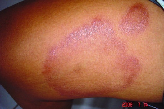
Caption: FIGURE 184-7 Type 1 leprosy reaction. Increased inflammation of existing lesions. (From Dr. W. H. van Brakel, with permission from NLR.) This reaction occurs mostly during multidrug therapy but can also develop in untreated patients. Evanescent, pink-to-red, maculopapular, papular, nodular, or plaque lesions suddenly appear and are usually accompanied by constitutional symptoms like malaise and fever, with 1. or without painful swelling in the joints (Fig. 184-8). These crops of skin lesions present on the outer aspects of the thighs, legs, and face. They are painful or tender and warm, blanch with light finger pres- — Figure 184-7: Type 1 leprosy reaction showing increased inflammation of existing lesions with acute swelling and redness.
Figure 8
Caption: FIGURE 184-3 Tuberculoid (TT) leprosy. Hypopigmented macular lesion with a well- FIGURE 184-4 Borderline tuberculoid (BT) leprosy. Macular lesion with irregular, defined edge and loss of fine-touch sensation. (From Dr. H. K. Kar, with permission.) moderately defined edge and satellite lesion, with loss of sensation. (From Dr. W. H. van Brakel, with permission from NLR.) and face, with mild to moderate impairment of touch and/or thermal sensations. There is no thickening of the corresponding cutaneous Mid-Borderline (BB) Leprosy This form of leprosy is unstable. and peripheral nerves. IL is often, but not always, the first clinical Many cases downgrade toward BL and LL disease, especially if not sign of leprosy. This type either heals spontaneously or progresses to treated. There are multiple plaque lesions and, not infrequently, macu- a determinate form of the disease (TT, BT, BB, BL, or LL), depending lar lesions; the lesions are of various shapes and sizes, are bilateral, and on CMI status. usually occur in a more or less symmetrical distribution. In annular — Figure 184-8: Type 2 leprosy reaction (erythema nodosum leprosum) showing evanescent, pink-to-red, maculopapular, nodular lesions with constitutional symptoms.
Generated from Harrison's Principles of Internal Medicine, 22nd Edition.