Gastrointestinal Endoscopy
Chapter 333 | Harrison's 22e · Part 10 – Gastrointestinal Disorders
Detailed clinical reference synthesised from Harrison's Principles of Internal Medicine, 22nd Edition
🔑 Key Clinical Points
- See source text for full details
📑 Table of Contents
📋 Figures in This Chapter
RAW CONTENT
[PAGE 2464] 2464 PART 10 Disorders of the Gastrointestinal System for nausea in first-trimester pregnancy. Peppermint oil and caraway acute cholecystitis in patients unable to undergo cholecystectomy. seed oil products and herbal preparations such as STW 5 (a nine- Transjugular intrahepatic portosystemic shunts are performed for herb mixture) are useful for functional dyspepsia and IBS. Low- variceal hemorrhage not amenable to endoscopic therapy. Litho- potency pancreatic enzyme preparations are sold as digestive aids tripsy is rarely performed to fragment gallstones in patients who but have little evidence to support their efficacy. are not surgical candidates. Radiologic approaches are often chosen over endoscopy for gastroenterostomy placement. Radiographic THERAPIES TARGETING GUT DYSBIOSIS assistance is sometimes needed for placement of central venous Antibiotics are prescribed to treat H. pylori–induced ulcers, infec- catheters for parenteral nutrition. tious diarrhea, complicated diverticulitis, and small intestinal SURGERY bacterial overgrowth. Some cases of diarrhea-predominant IBS respond to the nonabsorbable antibiotic rifaximin. Probiotics con- Roles of surgery in GI conditions include disease cure, symptom taining bacterial cultures and prebiotics that selectively nourish control, maintenance of nutrition, and palliation of unresectable nonnoxious commensal bacteria have been given as adjunctive neoplasm. Surgery cures medication-unresponsive ulcerative coli- therapy for infectious diarrhea and IBS, with limited evidence of tis, diverticulitis, cholecystitis, appendicitis, and intraabdominal efficacy. Postbiotics are metabolites made by probiotic organisms abscess, but only reduces symptoms and treats complications in that inhibit pathogenic luminal bacteria. Transplanting donor feces Crohn’s disease. Surgery is performed for ulcer complications like into the colon by colonoscopy or enema is effective treatment for bleeding, obstruction, or perforation and intestinal obstructions recurrent Clostridioides difficile colitis. Commercial oral and rectal that persist after conservative care. Gastroesophageal fundoplica- microbiota-based products have been approved or are in develop- tion is performed for refractory acid reflux. Acid exposure time ment for this indication. on pH testing helps select candidates for fundoplication. Achalasia responds to operations to reduce lower esophageal sphincter tone. THERAPEUTIC ENDOSCOPY Operations for motor disorders include implanted electrical stimu- In addition to its diagnostic role, endoscopy has numerous thera- lators for gastroparesis and electrical devices and artificial sphinc- peutic capabilities. Cautery techniques and injection of vasocon- ters for fecal incontinence. Surgery can place a jejunostomy for strictor substances can stop hemorrhage from ulcers and vascular long-term enteral feedings. Other common indications for surgery malformations. Endoscopically placed clips can occlude arterial include hernias, hemorrhoids, and nonhealing anal fissures. bleeding sites, while hemostatic powder sprays can stop brisk per- PSYCHOLOGICAL APPROACHES AND PHYSICAL THERAPY sistent GI bleeding. Endoscopic encirclement of esophageal varices and hemorrhoids with constricting bands stops hemorrhage from Psychological therapies, including psychotherapy, cognitive behav- these sites. Bleeding gastric varices can be injected with thrombin or ioral therapy, and hypnosis, show efficacy in DGBIs and are most cyanoacrylate. Endoscopy can remove polyps in the stomach, small beneficial for patients with significant psychological dysfunction. bowel, or colon. Decompressive colonoscopy withdraws excess Behavioral therapists provide instruction in diaphragmatic breath- gas in some cases of acute colonic pseudoobstruction. Endoscopic ing for belching or rumination. Biofeedback methods administered mucosal resection, submucosal dissection, and radiofrequency by physical therapists can treat refractory fecal incontinence or techniques ablate some cases of Barrett’s esophagus with dysplasia constipation secondary to dyssynergia. and resect superficial cancers or subepithelial tumors elsewhere in the gut. Obstructions of the GI lumen and pancreaticobiliary tree ■ FURTHER READING are relieved by endoscopic dilation or placing plastic or expandable Benech N et al: Update on microbiota-derived therapies for recurrent metal stents. Endoscopic sphincterotomy of the ampulla of Vater Clostridioides difficile infections. Clin Microbiol Infect 30:462, 2024. treats choledocholithiasis. Cholangioscopy can facilitate stone lith- Gergely M et al: Management of refractory inflammatory bowel otripsy in the common bile duct, ablation of small ductal tumors, disease. Curr Opin Gastroenterol 38:347, 2022. and placement of gallbladder stents to facilitate drainage in nonop- Hossain B et al: Prevalence and impact of gastrointestinal manifesta- erative candidates. Interventional EUS is used for pancreatic cyst tions in COVID-19 patients: A systematic review. J Community Hosp gastrostomy using lumen-apposing metal stents, pancreatic necro- Intern Med Perspect 13:39, 2023. sectomy, and placement of fiducial markers to direct pancreatic and Jain S et al: Optimal strategies for colorectal cancer screening. Curr rectal radiotherapy. EUS also can facilitate endoscopic access to the Treat Options Oncol 23:474, 2022. excluded distal stomach in patients who have undergone bariatric Orpen-Palmer J et al: Update on the management of upper gastroin- gastric bypass surgery using similar stents so that ERCP can be testinal bleeding. BMJ Med 1:e000202, 2022. done for pancreaticobiliary conditions. EUS-directed stent place- Shakir SM et al: Updates to the diagnosis and clinical management of ment can manage postsurgical stenoses after pancreatic resection. Helicobacter pylori infections. Clin Chem 69:869, 2023. Endoscopy is used to insert gastric feeding tubes. Peroral endo- scopic myotomy is performed on the lower esophageal sphincter in achalasia, the pylorus in gastroparesis, and the upper esophageal sphincter for Zenker’s diverticulum. Endoscopic treatments for acid reflux including transoral incisionless fundoplication have been developed as potential alternatives to surgery. Endoscopic bariatric methods, including intragastric balloons, sleeve gastroplasty, and duodenal resurfacing and diversion, have been devised. 333 Gastrointestinal INTERVENTIONAL RADIOLOGY Endoscopy Interventional radiology techniques offer benefits in selected set- tings. Angiographic embolization or vasoconstriction decreases Louis Michel Wong Kee Song, Vinay bleeding from gut sites not amenable to endoscopic intervention. Chandrasekhara, Mark Topazian Angiographic embolectomy, stent placement, and thrombolysis also manage mesenteric ischemia. Dilatation or stenting under fluoroscopic guidance relieves luminal strictures. Contrast enemas can reduce colon volvulus. CT- and ultrasound-directed drainage of Gastrointestinal endoscopy has been attempted for over 200 years, abdominal fluid collections can obviate the need for surgery. Per- but the introduction of semirigid and flexible gastroscopes in the cutaneous transhepatic cholangiography relieves biliary obstruction mid-twentieth century marked the dawn of the modern endoscopic when ERCP is contraindicated. Percutaneous cholecystostomy treats era. Since then, rapid advances in endoscopic technology have led to
[PAGE 2465] Gastrointestinal Endoscopy 2465 CHAPTER 333 FIGURE 333-1 Gastrointestinal endoscope. Shown here is a conventional colonoscope with control knobs for tip deflection, push buttons for suction and A air insufflation (single arrows), and a working channel for passage of accessories (double arrows). dramatic changes in the diagnosis and treatment of many digestive diseases. Innovative endoscopic devices and new endoscopic treatment modalities continue to expand the use of endoscopy in patient care. Flexible endoscop
Flowcharts & Algorithms
Reproduced from Harrison's 22nd Edition.
Flowchart 1

Caption: FIGURE 333-58 Endoscopic management of an esophagogastric anastomotic stricture. A. electroincision of stricture. C. Improvement in luminal opening after therapy.
Figures & Illustrations
Reproduced from Harrison's 22nd Edition.
Figure 1

Caption: FIGURE 333-11 Ulcerated colon adenocarcinoma narrowing the colonic lumen.
Figure 2

Caption: FIGURE 333-18 Endoscopic diagnosis, staging, and palliation of hilar obstructive jaundice demonstrates a malignant-appearing stricture of the biliary demonstrating a stricture with dilated, tortuous vessels with a malignant appearance. C. the submucosa of the bile duct wall (arrow). (Image courtesy of Dr. Thomas Smyrk.) D. posterior, and left systems relieves the biliary obstruction.
Figure 3

Caption: FIGURE 333-11 Ulcerated colon adenocarcinoma narrowing the colonic lumen.
Figure 4

Caption: FIGURE 333-19 Bile leak. A. Site of leak (arrow) from the cystic duct after laparoscopic into the gallbladder fossa (arrow).
Figure 5

Caption: FIGURE 333-16 Endoscopic retrograde cholangiopancreatography (ERCP) for bile duct stones. A. Faceted bile duct stones are demonstrated in the common bile duct and common hepatic duct. B. After endoscopic sphincterotomy, the stones are extracted with a stone extraction balloon.
Figure 6

Caption: FIGURE 333-16 Endoscopic retrograde cholangiopancreatography (ERCP) for bile duct stones. A. Faceted bile duct stones are demonstrated in the common bile duct and common hepatic duct. B. After endoscopic sphincterotomy, the stones are extracted with a stone extraction balloon.
Figure 7

Caption: FIGURE 333-18 Endoscopic diagnosis, staging, and palliation of hilar obstructive jaundice demonstrates a malignant-appearing stricture of the biliary demonstrating a stricture with dilated, tortuous vessels with a malignant appearance. C. the submucosa of the bile duct wall (arrow). (Image courtesy of Dr. Thomas Smyrk.) D. posterior, and left systems relieves the biliary obstruction.
Figure 8

Caption: FIGURE 333-18 Endoscopic diagnosis, staging, and palliation of hilar obstructive jaundice demonstrates a malignant-appearing stricture of the biliary demonstrating a stricture with dilated, tortuous vessels with a malignant appearance. C. the submucosa of the bile duct wall (arrow). (Image courtesy of Dr. Thomas Smyrk.) D. posterior, and left systems relieves the biliary obstruction.
Figure 9

Caption: FIGURE 333-19 Bile leak. A. Site of leak (arrow) from the cystic duct after laparoscopic into the gallbladder fossa (arrow).
Figure 10

Caption: FIGURE 333-17 Endoscopic sphincterotomy. A. A normal-appearing ampulla of Vater duct stones are extracted with a balloon catheter. endoscope have been used for many years to treat bleeding lesions, and the development of larger over-the-scope clips has facilitated endoscopic closure of gastrointestinal fistulas and perforations not previously amenable to endoscopic therapy (Video V5-7). Endoscopic suturing can be used to close some perforations and large defects (Fig. 333-27), anastomotic leaks, and fistulas. Endoscopic suturing may also be used to prevent stent migration (Fig. 333-28, Video V5-8) and to
Figure 11

Caption: FIGURE 333-18 Endoscopic diagnosis, staging, and palliation of hilar obstructive jaundice demonstrates a malignant-appearing stricture of the biliary demonstrating a stricture with dilated, tortuous vessels with a malignant appearance. C. the submucosa of the bile duct wall (arrow). (Image courtesy of Dr. Thomas Smyrk.) D. posterior, and left systems relieves the biliary obstruction.
Figure 12

Caption: FIGURE 333-17 Endoscopic sphincterotomy. A. A normal-appearing ampulla of Vater duct stones are extracted with a balloon catheter. endoscope have been used for many years to treat bleeding lesions, and the development of larger over-the-scope clips has facilitated endoscopic closure of gastrointestinal fistulas and perforations not previously amenable to endoscopic therapy (Video V5-7). Endoscopic suturing can be used to close some perforations and large defects (Fig. 333-27), anastomotic leaks, and fistulas. Endoscopic suturing may also be used to prevent stent migration (Fig. 333-28, Video V5-8) and to
Figure 13

Caption: FIGURE 333-17 Endoscopic sphincterotomy. A. A normal-appearing ampulla of Vater duct stones are extracted with a balloon catheter. endoscope have been used for many years to treat bleeding lesions, and the development of larger over-the-scope clips has facilitated endoscopic closure of gastrointestinal fistulas and perforations not previously amenable to endoscopic therapy (Video V5-7). Endoscopic suturing can be used to close some perforations and large defects (Fig. 333-27), anastomotic leaks, and fistulas. Endoscopic suturing may also be used to prevent stent migration (Fig. 333-28, Video V5-8) and to
Figure 14

Caption: FIGURE 333-31 (Continued)
Figure 15

Caption: FIGURE 333-41 Gastrointestinal vascular ectasias. A. Gastric antral vascular ectasia ectasias. B. Cecal vascular ectasia. C. Radiation-induced vascular ectasias of the rectum
Figure 16

Caption: FIGURE 333-41 Gastrointestinal vascular ectasias. A. Gastric antral vascular ectasia ectasias. B. Cecal vascular ectasia. C. Radiation-induced vascular ectasias of the rectum
Figure 17

Caption: FIGURE 333-54 Schatzki’s ring at the gastroesophageal junction.
Figure 18

Caption: FIGURE 333-30 Buried bumper syndrome. A. Migration of the internal disk bumper of B. Close-up view of the disk bumper (arrow) buried in the gastric wall.
Figure 19

Caption: FIGURE 333-9 Causes of colitis. A. Chronic ulcerative colitis with diffuse inflammation. B. adherent pseudomembranes. D. Ischemic colitis with patchy mucosal edema,
Figure 20

Caption: FIGURE 333-30 Buried bumper syndrome. A. Migration of the internal disk bumper of B. Close-up view of the disk bumper (arrow) buried in the gastric wall.
Figure 21

Caption: FIGURE 333-3 (Continued)
Figure 22

Caption: FIGURE 333-54 Schatzki’s ring at the gastroesophageal junction.
Figure 23

Caption: FIGURE 333-9 Causes of colitis. A. Chronic ulcerative colitis with diffuse inflammation. B. adherent pseudomembranes. D. Ischemic colitis with patchy mucosal edema,
Figure 24

Caption: FIGURE 333-9 Causes of colitis. A. Chronic ulcerative colitis with diffuse inflammation. B. adherent pseudomembranes. D. Ischemic colitis with patchy mucosal edema,
Figure 25

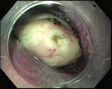
Caption: FIGURE 333-44 Esophageal food impaction. Meat bolus impacted in the distal esophagus.
Figure 26

Caption: FIGURE 333-30 Buried bumper syndrome. A. Migration of the internal disk bumper of B. Close-up view of the disk bumper (arrow) buried in the gastric wall.
Figure 27

Caption: FIGURE 333-39 (Continued)
Figure 28

Caption: FIGURE 333-3 (Continued)
Figure 29

Caption: FIGURE 333-33 Contact coagulation for ulcer hemostasis. A. Duodenal ulcer with a C. Obliteration of the treated vessel (arrow).
Figure 30

Caption: Gastrointestinal Endoscopy FIGURE 333-35 Over-the-scope clip placement for ulcer hemostasis. A. Pyloric channel ulcer with visible vessel (arrow). B. Hemostasis secured following placement of an over-the-scope clip. in ~50% of patients. Removable, fully covered lumen-apposing metal stents (LAMS) may also be used to treat benign pyloric stenosis (Video V5-16). Malignant gastric outlet obstruction can be relieved with endoscopically placed expandable stents across the obstruction in patients with inoperable malignancy (Video V5-17) or by EUS-guided B
Figure 31

Caption: FIGURE 333-9 Causes of colitis. A. Chronic ulcerative colitis with diffuse inflammation. B. adherent pseudomembranes. D. Ischemic colitis with patchy mucosal edema,
Figure 32

Caption: FIGURE 333-33 Contact coagulation for ulcer hemostasis. A. Duodenal ulcer with a C. Obliteration of the treated vessel (arrow).
Figure 33

Caption: Gastrointestinal Endoscopy FIGURE 333-35 Over-the-scope clip placement for ulcer hemostasis. A. Pyloric channel ulcer with visible vessel (arrow). B. Hemostasis secured following placement of an over-the-scope clip. in ~50% of patients. Removable, fully covered lumen-apposing metal stents (LAMS) may also be used to treat benign pyloric stenosis (Video V5-16). Malignant gastric outlet obstruction can be relieved with endoscopically placed expandable stents across the obstruction in patients with inoperable malignancy (Video V5-17) or by EUS-guided B
Figure 34

Caption: FIGURE 333-3 Normal upper endoscopic examination. A. Esophagus. B. G. Duodenal bulb. H. Second portion of the duodenum.
Figure 35

Caption: FIGURE 333-3 Normal upper endoscopic examination. A. Esophagus. B. G. Duodenal bulb. H. Second portion of the duodenum.
Figure 36

Caption: FIGURE 333-3 Normal upper endoscopic examination. A. Esophagus. B. G. Duodenal bulb. H. Second portion of the duodenum.
Figure 37

Caption: FIGURE 333-6 Barrett’s esophagus. A. Salmon-colored Barrett’s mucosa extending nodule (arrow) identified during endoscopic surveillance. C. Histologic finding of the esophageal submucosa (arrow). D. Barrett’s esophagus with locally advanced
Figure 38

Caption: FIGURE 333-6 Barrett’s esophagus. A. Salmon-colored Barrett’s mucosa extending nodule (arrow) identified during endoscopic surveillance. C. Histologic finding of the esophageal submucosa (arrow). D. Barrett’s esophagus with locally advanced
Figure 39

Caption: FIGURE 333-8 Normal colonoscopic examination. A. Cecum with view of appendiceal
Figure 40

Caption: FIGURE 333-8 Normal colonoscopic examination. A. Cecum with view of appendiceal
Figure 41

Caption: FIGURE 333-8 Normal colonoscopic examination. A. Cecum with view of appendiceal
Figure 42

Caption: FIGURE 333-8 Normal colonoscopic examination. A. Cecum with view of appendiceal
Figure 43

Caption: FIGURE 333-24 Endoscopic full-thickness resection (EFTR) of a gastrointestinal stromal from the fourth layer (muscularis propria) at endoscopic ultrasound. C. Full-thickness
Figure 44

Caption: FIGURE 333-38 Gastric varices. A. Large gastric fundal varices with stigmata of varix. C. Obliterated varix following glue injection on endoscopic follow-up at 1 month
Figure 45

Caption: FIGURE 333-42 Colonic diverticula. A
Figure 46

Caption: FIGURE 333-63 Small-bowel vascular ectasia. A. Actively bleeding mid-jejunal vascular with argon plasma coagulation (APC). C. Hemostasis secured following APC.
Figure 47

Caption: FIGURE 333-54 Schatzki’s ring at the gastroesophageal junction.
Figure 48

Caption: FIGURE 333-31 (Continued)
Figure 49

Caption: FIGURE 333-31 (Continued)
Figure 50

Caption: FIGURE 333-31 (Continued)
Figure 51

Caption: FIGURE 333-39 (Continued)
Figure 52

Caption: FIGURE 333-42 Colonic diverticula. A
Figure 53

Caption: FIGURE 333-48 Acute colonic pseudoobstruction. A. Acute colonic dilation occurring in a patient soon after knee surgery. B. Colonoscopic placement of decompression tube with marked improvement in colonic dilation.
Figure 54

Caption: FIGURE 333-48 Acute colonic pseudoobstruction. A. Acute colonic dilation occurring in a patient soon after knee surgery. B. Colonoscopic placement of decompression tube with marked improvement in colonic dilation.
Figure 55

Caption: FIGURE 333-3 (Continued)
Figure 56

Caption: FIGURE 333-3 (Continued)
Figure 57

Caption: FIGURE 333-11 Ulcerated colon adenocarcinoma narrowing the colonic lumen.
Figure 58

Caption: FIGURE 333-25 (Continued)
Figure 59

Caption: FIGURE 333-38 Gastric varices. A. Large gastric fundal varices with stigmata of varix. C. Obliterated varix following glue injection on endoscopic follow-up at 1 month
Figure 60

Caption: FIGURE 333-39 (Continued)
Figure 61

Caption: FIGURE 333-44 Esophageal food impaction. Meat bolus impacted in the distal esophagus.
Figure 62

Caption: FIGURE 333-1 Gastrointestinal endoscope. Shown here is a conventional colonoscope with control knobs for tip deflection, push buttons for suction and A air insufflation (single arrows), and a working channel for passage of accessories (double arrows).
Figure 63

Caption: FIGURE 333-1 Gastrointestinal endoscope. Shown here is a conventional colonoscope with control knobs for tip deflection, push buttons for suction and A air insufflation (single arrows), and a working channel for passage of accessories (double arrows).
Figure 64

Caption: FIGURE 333-26 (Continued)
Figure 65

Caption: FIGURE 333-51 Causes of esophagitis. A. Severe reflux esophagitis with mucosal esophagitis with target-type shallow ulcerations. D. Candida esophagitis with white
Figure 66

Caption: FIGURE 333-3 Normal upper endoscopic examination. A. Esophagus. B. G. Duodenal bulb. H. Second portion of the duodenum.
Figure 67

Caption: FIGURE 333-3 Normal upper endoscopic examination. A. Esophagus. B. G. Duodenal bulb. H. Second portion of the duodenum.
Figure 68

Caption: FIGURE 333-3 Normal upper endoscopic examination. A. Esophagus. B. G. Duodenal bulb. H. Second portion of the duodenum.
Figure 69

Caption: FIGURE 333-3 (Continued)
Figure 70

Caption: FIGURE 333-3 (Continued)
Figure 71

Caption: FIGURE 333-25 (Continued)
Figure 72

Caption: FIGURE 333-25 (Continued)
Figure 73

Caption: FIGURE 333-38 Gastric varices. A. Large gastric fundal varices with stigmata of varix. C. Obliterated varix following glue injection on endoscopic follow-up at 1 month
Figure 74

Caption: FIGURE 333-24 Endoscopic full-thickness resection (EFTR) of a gastrointestinal stromal from the fourth layer (muscularis propria) at endoscopic ultrasound. C. Full-thickness
Figure 75

Caption: FIGURE 333-26 (Continued)
Figure 76

Caption: FIGURE 333-33 Contact coagulation for ulcer hemostasis. A. Duodenal ulcer with a C. Obliteration of the treated vessel (arrow).
Figure 77

Caption: FIGURE 333-42 Colonic diverticula. A
Figure 78

Caption: FIGURE 333-56 (Continued)
Figure 79

Caption: FIGURE 333-25 (Continued)
Figure 80

Caption: FIGURE 333-38 Gastric varices. A. Large gastric fundal varices with stigmata of varix. C. Obliterated varix following glue injection on endoscopic follow-up at 1 month
Figure 81

Caption: FIGURE 333-38 Gastric varices. A. Large gastric fundal varices with stigmata of varix. C. Obliterated varix following glue injection on endoscopic follow-up at 1 month
Figure 82

Caption: FIGURE 333-41 Gastrointestinal vascular ectasias. A. Gastric antral vascular ectasia ectasias. B. Cecal vascular ectasia. C. Radiation-induced vascular ectasias of the rectum
Figure 83

Caption: FIGURE 333-52 (Continued)
Figure 84

Caption: FIGURE 333-16 Endoscopic retrograde cholangiopancreatography (ERCP) for bile duct stones. A. Faceted bile duct stones are demonstrated in the common bile duct and common hepatic duct. B. After endoscopic sphincterotomy, the stones are extracted with a stone extraction balloon.
Figure 85

Caption: FIGURE 333-23 Peroral endoscopic tumorectomy (POET). A. Mid-esophageal lesion. C. Submucosal dissection and tunneling to the site of the lesion. D. Dissection of through the muscularis propria. F. Mucosotomy site. G. Closure of mucosotomy site with
Figure 86

Caption: FIGURE 333-25 (Continued)
Figure 87

Caption: FIGURE 333-54 Schatzki’s ring at the gastroesophageal junction.
Figure 88

Caption: FIGURE 333-26 (Continued)
Figure 89

Caption: FIGURE 333-42 Colonic diverticula. A
Figure 90

Caption: FIGURE 333-51 Causes of esophagitis. A. Severe reflux esophagitis with mucosal esophagitis with target-type shallow ulcerations. D. Candida esophagitis with white
Figure 91

Caption: FIGURE 333-52 (Continued)
Figure 92

Caption: FIGURE 333-52 (Continued)
Figure 93

Caption: FIGURE 333-56 (Continued)
Figure 94

Caption: FIGURE 333-63 Small-bowel vascular ectasia. A. Actively bleeding mid-jejunal vascular with argon plasma coagulation (APC). C. Hemostasis secured following APC.
Figure 95

Caption: FIGURE 333-30 Buried bumper syndrome. A. Migration of the internal disk bumper of B. Close-up view of the disk bumper (arrow) buried in the gastric wall.
Figure 96

Caption: FIGURE 333-52 (Continued)
Figure 97

Caption: FIGURE 333-63 Small-bowel vascular ectasia. A. Actively bleeding mid-jejunal vascular with argon plasma coagulation (APC). C. Hemostasis secured following APC.
Figure 98

Caption: FIGURE 333-16 Endoscopic retrograde cholangiopancreatography (ERCP) for bile duct stones. A. Faceted bile duct stones are demonstrated in the common bile duct and common hepatic duct. B. After endoscopic sphincterotomy, the stones are extracted with a stone extraction balloon.
Figure 99

Caption: FIGURE 333-63 Small-bowel vascular ectasia. A. Actively bleeding mid-jejunal vascular with argon plasma coagulation (APC). C. Hemostasis secured following APC.
Figure 100

Caption: FIGURE 333-16 Endoscopic retrograde cholangiopancreatography (ERCP) for bile duct stones. A. Faceted bile duct stones are demonstrated in the common bile duct and common hepatic duct. B. After endoscopic sphincterotomy, the stones are extracted with a stone extraction balloon.
Figure 101

Caption: FIGURE 333-1 Gastrointestinal endoscope. Shown here is a conventional colonoscope with control knobs for tip deflection, push buttons for suction and A air insufflation (single arrows), and a working channel for passage of accessories (double arrows).
Figure 102

Caption: FIGURE 333-49 Obstructing colonic carcinoma. A. Colonic adenocarcinoma causing a self-expandable metal stent. C. Radiograph of expanded stent across the obstructing
Figure 103

Caption: FIGURE 333-49 Obstructing colonic carcinoma. A. Colonic adenocarcinoma causing a self-expandable metal stent. C. Radiograph of expanded stent across the obstructing
Figure 104

Caption: FIGURE 333-19 Bile leak. A. Site of leak (arrow) from the cystic duct after laparoscopic into the gallbladder fossa (arrow).
Figure 105

Caption: FIGURE 333-49 Obstructing colonic carcinoma. A. Colonic adenocarcinoma causing a self-expandable metal stent. C. Radiograph of expanded stent across the obstructing
Figure 106

Caption: FIGURE 333-60 Placement of biliary and duodenal self-expanding metal stents (SEMS) for obstruction caused by pancreatic cancer. A. Endoscopic retrograde cholangiopancreatography (ERCP) demonstrates a distal bile duct stricture (arrow). B. A biliary SEMS is placed. C. Contrast injection demonstrates a duodenal stricture (arrow). D. Biliary and duodenal SEMS in place.
Figure 107

Caption: FIGURE 333-60 Placement of biliary and duodenal self-expanding metal stents (SEMS) for obstruction caused by pancreatic cancer. A. Endoscopic retrograde cholangiopancreatography (ERCP) demonstrates a distal bile duct stricture (arrow). B. A biliary SEMS is placed. C. Contrast injection demonstrates a duodenal stricture (arrow). D. Biliary and duodenal SEMS in place.
Figure 108

Caption: FIGURE 333-9 Causes of colitis. A. Chronic ulcerative colitis with diffuse inflammation. B. adherent pseudomembranes. D. Ischemic colitis with patchy mucosal edema,
Figure 109

Caption: FIGURE 333-19 Bile leak. A. Site of leak (arrow) from the cystic duct after laparoscopic into the gallbladder fossa (arrow).
Figure 110

Caption: FIGURE 333-60 Placement of biliary and duodenal self-expanding metal stents (SEMS) for obstruction caused by pancreatic cancer. A. Endoscopic retrograde cholangiopancreatography (ERCP) demonstrates a distal bile duct stricture (arrow). B. A biliary SEMS is placed. C. Contrast injection demonstrates a duodenal stricture (arrow). D. Biliary and duodenal SEMS in place.
Figure 111

Caption: FIGURE 333-48 Acute colonic pseudoobstruction. A. Acute colonic dilation occurring in a patient soon after knee surgery. B. Colonoscopic placement of decompression tube with marked improvement in colonic dilation.
Figure 112

Caption: FIGURE 333-60 Placement of biliary and duodenal self-expanding metal stents (SEMS) for obstruction caused by pancreatic cancer. A. Endoscopic retrograde cholangiopancreatography (ERCP) demonstrates a distal bile duct stricture (arrow). B. A biliary SEMS is placed. C. Contrast injection demonstrates a duodenal stricture (arrow). D. Biliary and duodenal SEMS in place.
Figure 113

Caption: FIGURE 333-19 Bile leak. A. Site of leak (arrow) from the cystic duct after laparoscopic into the gallbladder fossa (arrow).
Figure 114

Caption: FIGURE 333-26 (Continued)
Figure 115

Caption: FIGURE 333-27 (Continued)
Figure 116

Caption: FIGURE 333-27 (Continued)
Figure 117

Caption: FIGURE 333-27 (Continued)
Figure 118

Caption: FIGURE 333-27 (Continued)
Figure 119

Caption: FIGURE 333-49 Obstructing colonic carcinoma. A. Colonic adenocarcinoma causing a self-expandable metal stent. C. Radiograph of expanded stent across the obstructing
Figure 120

Caption: FIGURE 333-49 Obstructing colonic carcinoma. A. Colonic adenocarcinoma causing a self-expandable metal stent. C. Radiograph of expanded stent across the obstructing
Figure 121

Caption: FIGURE 333-19 Bile leak. A. Site of leak (arrow) from the cystic duct after laparoscopic into the gallbladder fossa (arrow).
Figure 122

Caption: FIGURE 333-49 Obstructing colonic carcinoma. A. Colonic adenocarcinoma causing a self-expandable metal stent. C. Radiograph of expanded stent across the obstructing
Figure 123

Caption: FIGURE 333-44 Esophageal food impaction. Meat bolus impacted in the distal esophagus.
Figure 124

Caption: FIGURE 333-44 Esophageal food impaction. Meat bolus impacted in the distal esophagus.
Figure 125

Caption: FIGURE 333-26 (Continued)
Figure 126

Caption: FIGURE 333-44 Esophageal food impaction. Meat bolus impacted in the distal esophagus.
Figure 127

Caption: FIGURE 333-9 Causes of colitis. A. Chronic ulcerative colitis with diffuse inflammation. B. adherent pseudomembranes. D. Ischemic colitis with patchy mucosal edema,
Figure 128

Caption: FIGURE 333-29 Bleeding from percutaneous endoscopic gastrostomy (PEG) tube internal disk bumper of the PEG tube revealed active bleeding from within the PEG tract.
Figure 129

Caption: FIGURE 333-29 Bleeding from percutaneous endoscopic gastrostomy (PEG) tube internal disk bumper of the PEG tube revealed active bleeding from within the PEG tract.
Figure 130

Caption: Gastrointestinal Endoscopy FIGURE 333-35 Over-the-scope clip placement for ulcer hemostasis. A. Pyloric channel ulcer with visible vessel (arrow). B. Hemostasis secured following placement of an over-the-scope clip. in ~50% of patients. Removable, fully covered lumen-apposing metal stents (LAMS) may also be used to treat benign pyloric stenosis (Video V5-16). Malignant gastric outlet obstruction can be relieved with endoscopically placed expandable stents across the obstruction in patients with inoperable malignancy (Video V5-17) or by EUS-guided B
Figure 131

Caption: FIGURE 333-27 (Continued)
Figure 132

Caption: FIGURE 333-23 Peroral endoscopic tumorectomy (POET). A. Mid-esophageal lesion. C. Submucosal dissection and tunneling to the site of the lesion. D. Dissection of through the muscularis propria. F. Mucosotomy site. G. Closure of mucosotomy site with
Figure 133

Caption: FIGURE 333-23 Peroral endoscopic tumorectomy (POET). A. Mid-esophageal lesion. C. Submucosal dissection and tunneling to the site of the lesion. D. Dissection of through the muscularis propria. F. Mucosotomy site. G. Closure of mucosotomy site with
Figure 134
Caption: FIGURE 333-23 Peroral endoscopic tumorectomy (POET). A. Mid-esophageal lesion. C. Submucosal dissection and tunneling to the site of the lesion. D. Dissection of through the muscularis propria. F. Mucosotomy site. G. Closure of mucosotomy site with
Figure 135

Caption: FIGURE 333-23 Peroral endoscopic tumorectomy (POET). A. Mid-esophageal lesion. C. Submucosal dissection and tunneling to the site of the lesion. D. Dissection of through the muscularis propria. F. Mucosotomy site. G. Closure of mucosotomy site with
Figure 136

Caption: FIGURE 333-23 Peroral endoscopic tumorectomy (POET). A. Mid-esophageal lesion. C. Submucosal dissection and tunneling to the site of the lesion. D. Dissection of through the muscularis propria. F. Mucosotomy site. G. Closure of mucosotomy site with
Figure 137

Caption: FIGURE 333-23 Peroral endoscopic tumorectomy (POET). A. Mid-esophageal lesion. C. Submucosal dissection and tunneling to the site of the lesion. D. Dissection of through the muscularis propria. F. Mucosotomy site. G. Closure of mucosotomy site with
Figure 138

Caption: FIGURE 333-23 Peroral endoscopic tumorectomy (POET). A. Mid-esophageal lesion. C. Submucosal dissection and tunneling to the site of the lesion. D. Dissection of through the muscularis propria. F. Mucosotomy site. G. Closure of mucosotomy site with
Figure 139

Caption: FIGURE 333-11 Ulcerated colon adenocarcinoma narrowing the colonic lumen.
Figure 140

Caption: FIGURE 333-11 Ulcerated colon adenocarcinoma narrowing the colonic lumen.
Figure 141

Caption: FIGURE 333-33 Contact coagulation for ulcer hemostasis. A. Duodenal ulcer with a C. Obliteration of the treated vessel (arrow).
Figure 142

Caption: FIGURE 333-33 Contact coagulation for ulcer hemostasis. A. Duodenal ulcer with a C. Obliteration of the treated vessel (arrow).
Figure 143

Caption: FIGURE 333-60 Placement of biliary and duodenal self-expanding metal stents (SEMS) for obstruction caused by pancreatic cancer. A. Endoscopic retrograde cholangiopancreatography (ERCP) demonstrates a distal bile duct stricture (arrow). B. A biliary SEMS is placed. C. Contrast injection demonstrates a duodenal stricture (arrow). D. Biliary and duodenal SEMS in place.
Figure 144

Caption: FIGURE 333-60 Placement of biliary and duodenal self-expanding metal stents (SEMS) for obstruction caused by pancreatic cancer. A. Endoscopic retrograde cholangiopancreatography (ERCP) demonstrates a distal bile duct stricture (arrow). B. A biliary SEMS is placed. C. Contrast injection demonstrates a duodenal stricture (arrow). D. Biliary and duodenal SEMS in place.
Figure 145

Caption: Gastrointestinal Endoscopy FIGURE 333-35 Over-the-scope clip placement for ulcer hemostasis. A. Pyloric channel ulcer with visible vessel (arrow). B. Hemostasis secured following placement of an over-the-scope clip. in ~50% of patients. Removable, fully covered lumen-apposing metal stents (LAMS) may also be used to treat benign pyloric stenosis (Video V5-16). Malignant gastric outlet obstruction can be relieved with endoscopically placed expandable stents across the obstruction in patients with inoperable malignancy (Video V5-17) or by EUS-guided B
Figure 146

Caption: FIGURE 333-11 Ulcerated colon adenocarcinoma narrowing the colonic lumen.
Figure 147

Caption: FIGURE 333-56 (Continued)
Figure 148

Caption: FIGURE 333-56 (Continued)
Figure 149

Caption: FIGURE 333-56 (Continued)
Figure 150

Caption: Gastrointestinal Endoscopy FIGURE 333-35 Over-the-scope clip placement for ulcer hemostasis. A. Pyloric channel ulcer with visible vessel (arrow). B. Hemostasis secured following placement of an over-the-scope clip. in ~50% of patients. Removable, fully covered lumen-apposing metal stents (LAMS) may also be used to treat benign pyloric stenosis (Video V5-16). Malignant gastric outlet obstruction can be relieved with endoscopically placed expandable stents across the obstruction in patients with inoperable malignancy (Video V5-17) or by EUS-guided B
Figure 151

Caption: FIGURE 333-24 Endoscopic full-thickness resection (EFTR) of a gastrointestinal stromal from the fourth layer (muscularis propria) at endoscopic ultrasound. C. Full-thickness
Figure 152

Caption: FIGURE 333-24 Endoscopic full-thickness resection (EFTR) of a gastrointestinal stromal from the fourth layer (muscularis propria) at endoscopic ultrasound. C. Full-thickness
Figure 153

Caption: FIGURE 333-24 Endoscopic full-thickness resection (EFTR) of a gastrointestinal stromal from the fourth layer (muscularis propria) at endoscopic ultrasound. C. Full-thickness
Figure 154

Caption: FIGURE 333-24 Endoscopic full-thickness resection (EFTR) of a gastrointestinal stromal from the fourth layer (muscularis propria) at endoscopic ultrasound. C. Full-thickness
Figure 155

Caption: FIGURE 333-27 (Continued)
Figure 156

Caption: FIGURE 333-6 Barrett’s esophagus. A. Salmon-colored Barrett’s mucosa extending nodule (arrow) identified during endoscopic surveillance. C. Histologic finding of the esophageal submucosa (arrow). D. Barrett’s esophagus with locally advanced
Figure 157

Caption: FIGURE 333-22 Peroral endoscopic myotomy (POEM) for achalasia. A. Dilated (LES) region. C. Mucosal incision (mucosotomy) 10 cm proximal to the LES. D. mucosotomy site into the submucosal space. E. Completion of submucosal tunnel to the site. G. Completion of myotomy to the cardia. H. Closure of mucosotomy site with clips. I.
Figure 158

Caption: FIGURE 333-22 Peroral endoscopic myotomy (POEM) for achalasia. A. Dilated (LES) region. C. Mucosal incision (mucosotomy) 10 cm proximal to the LES. D. mucosotomy site into the submucosal space. E. Completion of submucosal tunnel to the site. G. Completion of myotomy to the cardia. H. Closure of mucosotomy site with clips. I.
Figure 159

Caption: FIGURE 333-22 Peroral endoscopic myotomy (POEM) for achalasia. A. Dilated (LES) region. C. Mucosal incision (mucosotomy) 10 cm proximal to the LES. D. mucosotomy site into the submucosal space. E. Completion of submucosal tunnel to the site. G. Completion of myotomy to the cardia. H. Closure of mucosotomy site with clips. I.
Figure 160

Caption: FIGURE 333-22 Peroral endoscopic myotomy (POEM) for achalasia. A. Dilated (LES) region. C. Mucosal incision (mucosotomy) 10 cm proximal to the LES. D. mucosotomy site into the submucosal space. E. Completion of submucosal tunnel to the site. G. Completion of myotomy to the cardia. H. Closure of mucosotomy site with clips. I.
Figure 161

Caption: FIGURE 333-22 Peroral endoscopic myotomy (POEM) for achalasia. A. Dilated (LES) region. C. Mucosal incision (mucosotomy) 10 cm proximal to the LES. D. mucosotomy site into the submucosal space. E. Completion of submucosal tunnel to the site. G. Completion of myotomy to the cardia. H. Closure of mucosotomy site with clips. I.
Figure 162

Caption: FIGURE 333-22 Peroral endoscopic myotomy (POEM) for achalasia. A. Dilated (LES) region. C. Mucosal incision (mucosotomy) 10 cm proximal to the LES. D. mucosotomy site into the submucosal space. E. Completion of submucosal tunnel to the site. G. Completion of myotomy to the cardia. H. Closure of mucosotomy site with clips. I.
Figure 163

Caption: FIGURE 333-22 Peroral endoscopic myotomy (POEM) for achalasia. A. Dilated (LES) region. C. Mucosal incision (mucosotomy) 10 cm proximal to the LES. D. mucosotomy site into the submucosal space. E. Completion of submucosal tunnel to the site. G. Completion of myotomy to the cardia. H. Closure of mucosotomy site with clips. I.
Figure 164

Caption: FIGURE 333-22 Peroral endoscopic myotomy (POEM) for achalasia. A. Dilated (LES) region. C. Mucosal incision (mucosotomy) 10 cm proximal to the LES. D. mucosotomy site into the submucosal space. E. Completion of submucosal tunnel to the site. G. Completion of myotomy to the cardia. H. Closure of mucosotomy site with clips. I.
Figure 165

Caption: FIGURE 333-22 Peroral endoscopic myotomy (POEM) for achalasia. A. Dilated (LES) region. C. Mucosal incision (mucosotomy) 10 cm proximal to the LES. D. mucosotomy site into the submucosal space. E. Completion of submucosal tunnel to the site. G. Completion of myotomy to the cardia. H. Closure of mucosotomy site with clips. I.
Figure 166

Caption: FIGURE 333-6 Barrett’s esophagus. A. Salmon-colored Barrett’s mucosa extending nodule (arrow) identified during endoscopic surveillance. C. Histologic finding of the esophageal submucosa (arrow). D. Barrett’s esophagus with locally advanced
Figure 167

Caption: FIGURE 333-6 Barrett’s esophagus. A. Salmon-colored Barrett’s mucosa extending nodule (arrow) identified during endoscopic surveillance. C. Histologic finding of the esophageal submucosa (arrow). D. Barrett’s esophagus with locally advanced
Figure 168

Caption: FIGURE 333-48 Acute colonic pseudoobstruction. A. Acute colonic dilation occurring in a patient soon after knee surgery. B. Colonoscopic placement of decompression tube with marked improvement in colonic dilation.
Figure 169

Caption: FIGURE 333-48 Acute colonic pseudoobstruction. A. Acute colonic dilation occurring in a patient soon after knee surgery. B. Colonoscopic placement of decompression tube with marked improvement in colonic dilation.
Figure 170

Caption: FIGURE 333-51 Causes of esophagitis. A. Severe reflux esophagitis with mucosal esophagitis with target-type shallow ulcerations. D. Candida esophagitis with white
Figure 171

Caption: FIGURE 333-51 Causes of esophagitis. A. Severe reflux esophagitis with mucosal esophagitis with target-type shallow ulcerations. D. Candida esophagitis with white
Figure 172

Caption: FIGURE 333-51 Causes of esophagitis. A. Severe reflux esophagitis with mucosal esophagitis with target-type shallow ulcerations. D. Candida esophagitis with white
Figure 173

Caption: FIGURE 333-51 Causes of esophagitis. A. Severe reflux esophagitis with mucosal esophagitis with target-type shallow ulcerations. D. Candida esophagitis with white
Figure 174

Caption: FIGURE 333-6 Barrett’s esophagus. A. Salmon-colored Barrett’s mucosa extending nodule (arrow) identified during endoscopic surveillance. C. Histologic finding of the esophageal submucosa (arrow). D. Barrett’s esophagus with locally advanced
Generated from Harrison's Principles of Internal Medicine, 22nd Edition.