Multiple Sclerosis
Chapter 455 | Part 13: Neurologic Disorders · Part 13 – Neurologic Disorders
Detailed clinical reference synthesised from Harrison's Principles of Internal Medicine, 22nd Edition
🔑 Key Clinical Points
- MS is an autoimmune disease of the CNS characterized by chronic inflammation, demyelination, gliosis, and neuronal loss.
- Women are affected approximately three times more often than men; onset typically between 20 and 40 years.
- Clinical course ranges from relapsing-remitting (RRMS) to primary progressive (PPMS) to secondary progressive (SPMS).
- Diagnosis requires dissemination in time (two or more episodes) and space (two or more CNS locations).
- EBV infection is a near-universal prerequisite for MS development; history of infectious mononucleosis increases risk.
- HLA-DRB1*1501 is the strongest genetic susceptibility signal, accounting for ~10% of disease risk.
- Disability accumulation is driven by 'silent progression' (PIRA) independent of relapse activity in the treatment era.
- EDSS score of 4 or greater plus FSS motor score of 2 or greater supports diagnosis of SPMS.
- Heat sensitivity (Uhthoff's symptom) and Lhermitte's symptom are characteristic clinical features.
- B cell–based treatments and highly effective therapies have dramatically improved long-term outcomes.
📑 Table of Contents
- 1. DEFINITION & OVERVIEW
- 1.1 Disease Course Classifications
- 2. EPIDEMIOLOGY
- 3. ETIOLOGY & PATHOPHYSIOLOGY
- 3.1 Pathology
- 3.2 Physiology
- 4. CLINICAL FEATURES
- 4.1 Sensory Symptoms
- 4.2 Visual Symptoms
- 4.3 Motor Symptoms
- 4.4 Ancillary Symptoms
- 5. DIFFERENTIAL DIAGNOSIS
- 6. INVESTIGATIONS & DIAGNOSIS
- 6.1 Diagnostic Criteria
- 6.2 Scoring Systems
- 7. MANAGEMENT & TREATMENT
- 7.1 Therapeutic Goals
- 7.2 Treatment Classes
- 8. PROGNOSIS & COMPLICATIONS
- 9. SPECIAL CONSIDERATIONS
- 10. KEY PEARLS & CLINICAL TRAPS
- Figures & Illustrations
📋 Figures in This Chapter
1. DEFINITION & OVERVIEW
- Multiple sclerosis (MS) is an autoimmune disease of the central nervous system (CNS) characterized by chronic inflammation, demyelination, gliosis (plaques or scarring), and neuronal loss.
- The course can be relapsing or progressive.
- MS plaques typically develop at different times and in different CNS locations (i.e., MS is said to be disseminated in time and space).
- One million individuals in the United States, and millions worldwide, are affected.
- The clinical course is extremely variable, ranging from a relatively benign condition to a rapidly evolving and incapacitating disease requiring profound lifestyle adjustments.
- The past decade has seen tremendous progress in understanding basic disease mechanisms underlying MS and in developing highly effective therapies especially for the relapsing form of the disease.
- These advances have dramatically improved the long-term outcome for patients.
- Autopsy series identified MS in some individuals (~0.1% of cases) who were seemingly asymptomatic during life, and magnetic resonance imaging (MRI) scans obtained for unrelated reasons also showed evidence of asymptomatic MS, an incidental finding termed a radiologically isolated syndrome (RIS).
- Specific symptoms of MS are varied and reflect the location and severity of lesions within the CNS.
1.1 Disease Course Classifications
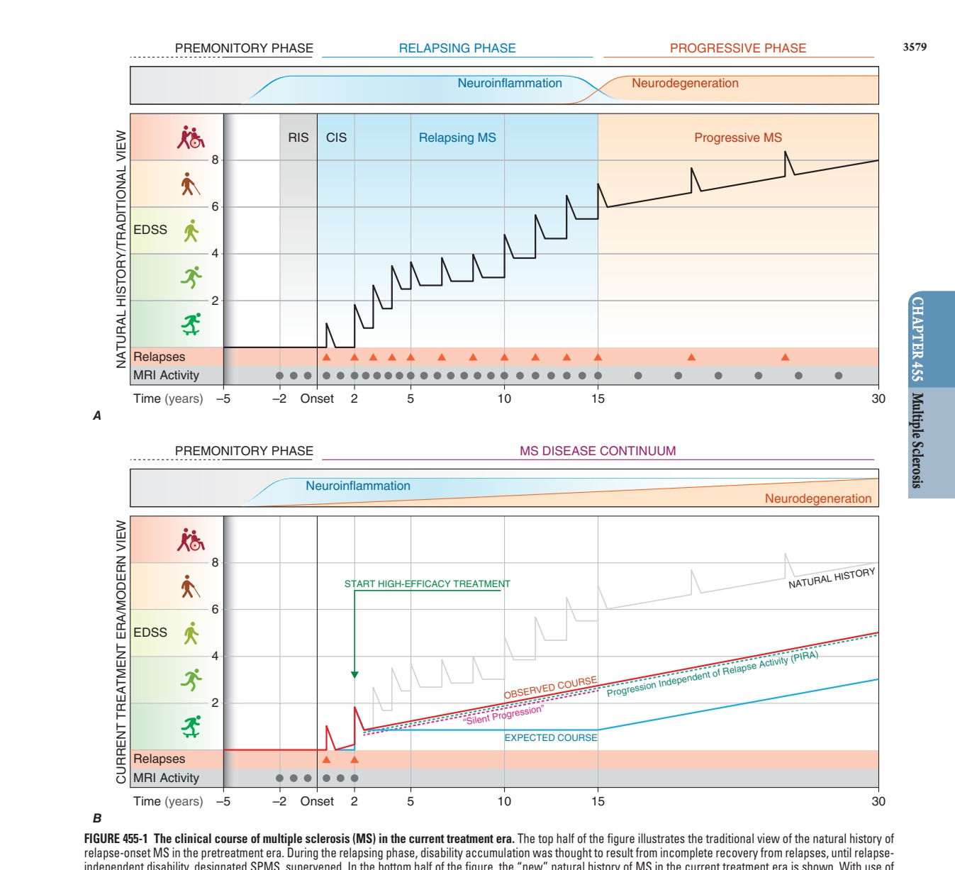
- In the traditional model of MS, the disease was considered to have three principal clinical forms, designated relapsing-remitting, secondary progressive, and primary progressive.
- More recently, these categories were supplanted by a unitary view of the disease, in which inflammation and neurodegeneration are present in most patients throughout the disease course.
- Nonetheless, from a clinical perspective, it is still often useful to apply the classical subtype scheme to assessment and management of patients.
-
- Relapsing-remitting or bout onset MS (RRMS):
- Accounts for 90% of MS cases.
- Characterized by discrete attacks of neurologic dysfunction that generally evolve over days to weeks (rarely over hours).
- In early MS, there is often substantial or complete recovery over the ensuing weeks to months.
- As attacks continue, recovery may be less evident.
- Between attacks, patients were earlier thought to be neurologically stable; however, it is now clear that most if not all patients with RRMS experience subtle 'silent' progression even when relapse-free.
-
- Secondary progressive MS (SPMS):
- Always begins as RRMS.
- At some point, the clinical course changes so that the patient experiences progressive deterioration in function unassociated with acute attacks.
- SPMS produces a greater amount of fixed neurologic course disability than RRMS.
- A practical definition for SPMS is a patient who has developed some level of permanent walking disability not due exclusively to relapses.
- The Extended Disability Status Score (EDSS) is a widely used measure of neurologic impairment in MS.
- An EDSS of 4 or greater, plus a Functional Status Scale (FSS) motor system score of 2 or greater, can support a diagnosis of SPMS.
- For a patient with RRMS, in the pretreatment era, the risk of developing SPMS was ~3% each year, meaning that the great majority of RRMS would ultimately evolve into SPMS.
- However, more recent case series have indicated a much lower rate of evolution to SPMS, estimated at <1% each year, likely due to widespread use of increasingly effective therapies for MS.
-
- Primary progressive MS (PPMS):
- Accounts for ~10% of cases.
- These patients do not experience attacks but rather steadily decline in function from disease onset.
- Compared to RRMS, the sex distribution is more even, the disease begins later in life (mean age ~40 years), and disability develops faster relative to the onset of the first clinical symptom.
- Despite these differences PPMS appears to represent the same underlying illness as RRMS and SPMS.
- Some PPMS patients experience relapses over the course of their illness.
- The term active progressive MS is used to categorize progressive MS patients (both SPMS and PPMS) who experience relapses or are found to have new lesions on serial MRI scans.
2. EPIDEMIOLOGY
- Geographic gradients are consistently observed in MS, with the highest prevalence generally found in temperate zones.
- In tropical regions, prevalence is often 10-fold to 20-fold less.
- A north-south gradient was observed in numerous national and regional studies, with decreasing rates as one moves equatorially.
- The prevalence of MS also increased steadily in several regions around the world over the past half-century, presumably reflecting the impact of some environmental shift, improved diagnosis, and/or a longer lifespan.
- This increase appears to have occurred to a greater degree in women than men and in nonwhite populations.
- In the United States, there is a slightly higher prevalence in white compared with black individuals, with lower estimates in Hispanics, followed by Asians.
3. ETIOLOGY & PATHOPHYSIOLOGY
- Multiple lines of evidence incriminate a role for infection with the Epstein-Barr virus (EBV) in MS.
- Individuals who have never been EBV infected (~5% of the population globally) have a very low MS risk, ~20-fold lower than in EBV-positive individuals.
- A history of infectious mononucleosis (associated with initial exposure to EBV during adolescence or later in life) increases risk more than twofold higher yet.
- Higher antibody titers to EBV nuclear antigens were repeatedly associated with MS risk.
- Studies from longitudinal biobank collections showed that serologic conversion to EBV is a near-universal prerequisite for development of MS.
- Following primary EBV infection, a lifelong infection is established in most individuals, with latent EBV exclusively present in very small numbers (~1:10^-6) of B lymphocytes.
- EBV-infected B cells were not consistently identified in the nervous system of MS patients.
- It is possible that ongoing lytic cycles by very few infected B cells residing within the CNS could produce bursts of inflammation and MS lesions; however, it is more likely that pathology could be triggered by B cell–mediated antigen presentation of EBV peptides that cross-react with MS autoantigens via molecular mimicry.
- A history of cigarette smoking is also associated with MS risk.
- Interestingly, in an animal model of MS, the lung was identified as a critical site for activation of pathogenic T lymphocytes responsible for autoimmune demyelination.
- Finally, vitamin D deficiency has been repeatedly associated with MS.
- Immunoregulatory effects of vitamin D could explain these apparent relationships.
- Exposure of the skin to ultraviolet B (UVB) radiation from the sun is essential for the biosynthesis of vitamin D, and this endogenous production is the most important source of vitamin D in most individuals.
- A diet rich in fatty fish represents another source of vitamin D.
- At higher latitudes, the amount of UVB radiation reaching the earth's surface is often insufficient, particularly during winter months, and consequently, low serum levels of vitamin D are frequent in temperate zones.
- The common practice to avoid direct sun exposure and the widespread use of sunblock would be expected to exacerbate any population-wide vitamin D deficiency.
- MS aggregates within some families, and adoption, half-sibling, twin, and spousal studies indicate that familial aggregation is primarily due to genetic factors.
- Importantly, family studies also support a contribution of environment, as fraternal twins of MS patients are at higher risk than nontwin siblings.
- Susceptibility to MS is polygenic, with each gene contributing a relatively small amount to overall risk.
- The strongest susceptibility signal genome-wide maps to the human leukocyte antigen (HLA)-DRB1 gene in the class II region of the major histocompatibility complex (MHC) and specifically to HLA-DRB1*1501 (formerly designated DR2).
- This association accounts for ~10% of the disease risk.
- This HLA association, first described in the early 1970s, suggests that MS, at its core, is an autoimmune disease.
- Whole-genome association studies have identified >230 other MS susceptibility variants, each of which individually has only a very small effect on MS risk.
- Many of these MS-associated genes have known roles in the adaptive and innate immune system, for example, the genes for the interleukin (IL) 7 receptor (CD127), IL-2 receptor (CD25), and T-cell costimulatory molecule LFA-3 (CD58).
- Some variants also influence susceptibility to other autoimmune diseases in addition to MS.
- The variants identified so far all lack specificity in the cerebral cortex, and diffusible factors from these lymphoid cells appear to mediate subpial cortical demyelination and neurodegeneration.
- Cux2-positive neurons in layer 2 and 3 of the neocortex appear to be particularly vulnerable.
- Neuronal and axonal death may result from glutamate-mediated excitotoxicity, oxidative injury, iron accumulation, and/or mitochondrial failure.
- In relapsing MS, inflammation is characterized by focal perivenular infiltration of lymphocytes and monocytes, BBB disruption, and active demyelination.
- By contrast, inflammation in progressive MS is more diffuse, with widespread microglial proliferation across large areas of white matter, accompanied by infiltration of CD8 T cells and plasma blasts/plasma cells.
- Reduced myelin staining and axonal injury ('dirty white matter') are associated with these chronic pathologies.
- Astrogliosis has long been known to be a prominent feature of MS pathology, and activated astrocytes likely contribute directly to neuronal and myelin injury.
- Ongoing inflammation occurs behind an intact BBB in many patients with progressive MS, possibly accounting for the failure of immunotherapies not capable of crossing the BBB to benefit patients with progressive MS.
- An autoimmune response directed against components of CNS myelin, and perhaps other neural elements as well, remains the cornerstone of current concepts of MS pathogenesis.
- However, specific antigenic targets in MS have never been conclusively identified.
- B cells are centrally involved in the development of demyelinating lesions, as evidenced by the efficacy of B cell–based treatments in all forms of MS.
- Clonally restricted populations of activated, antigen-experienced, memory B cells and plasma cells are present in MS lesions, in meningeal lymphoid follicle-like structures overlying the cerebral cortex, and in cerebrospinal fluid (CSF).
- They produce the oligoclonal immunoglobulins and increased antibody synthesis rates in the CSF long useful in the diagnosis of MS.
- Myelin-specific autoantibodies, some directed against an extracellular myelin protein, myelin oligodendrocyte glycoprotein (MOG), have been detected bound to degenerating myelin in MS plaques.
- However, many more antibodies derived from these B cells appear to be directed against a diverse array of ubiquitous intracellular proteins seemingly unrelated to MS pathogenesis.
- Furthermore, the specific targets are different in each patient.
- Therefore, although these highly restricted CNS antibodies are characteristic of MS, their role in disease remains uncertain.
- More likely, the antigen-presenting cell (APC) function of B cells explains their role in MS pathogenesis.
- Fragments of self-peptides derived from HLA-DR2 proteins themselves were found to bind intact DRB1*1501 molecules on B cells and serve as antigens for presentation to T cells.
- Memory CD4+ T cells derived from CSF responded to these self-peptides bound to DR2 molecules, and in some cases, these self-peptides were cross-reactive with several myelin antigens, as well as proteins derived from EBV, Akkermansia muciniphila (a commensal gut bacterium associated with dysbiosis in MS patients), and RAS guanyl-releasing protein 2 (RASGRP2), previously found to be a possible T-cell autoantigen in MS.
- Thus, MS-associated HLA proteins contain fragments that might trigger autoimmunity through molecular mimicry with viral, bacterial, or norm
3.1 Pathology
- Demyelination: New MS lesions begin with perivenular cuffing by inflammatory mononuclear cells, predominantly T cells and macrophages, which also infiltrate the surrounding white matter.
- At sites of inflammation, the blood-brain barrier (BBB) is disrupted, but unlike vasculitis, the vessel wall is preserved.
- At the leading edge of lesions, cytotoxic CD8 cells are found.
- Involvement of the humoral immune system is also evident; B lymphocytes infiltrate the nervous system, myelin-specific autoantibodies are present on degenerating myelin sheaths, and complement is activated.
- Sharply demarcated areas of demyelination are the pathologic hallmark of MS lesions, and evidence of myelin degeneration is found at the earliest time points of tissue injury.
- Although relative sparing of axons is typical, partial or total axonal destruction can also occur, especially within highly inflammatory lesions.
- In some lesions, surviving oligodendrocytes or those that differentiate from precursor cells partially remyelinate the surviving axons, producing so-called shadow plaques.
- However, in many lesions, oligodendrocyte precursor cells are present but fail to differentiate into mature myelin-producing cells.
- Therefore, promoting remyelination to protect axons remains an important therapeutic goal.
- As lesions evolve, there is prominent astrocytic proliferation (gliosis), and the term sclerosis refers to these gliotic plaques that have a rubbery or hardened texture at autopsy.
- Neurodegeneration: Cumulative axonal and neuronal loss is the most important contributor to irreversible neurologic disability and progressive symptoms.
- With paraplegia due to MS, as many as 70% of axons are ultimately lost from the lateral corticospinal (e.g., motor) tracts.
- Demyelination can reduce trophic support for axons, redistribute ion channels, and destabilize action potential membrane potentials.
- Axons can adapt initially to these injuries, but over time, distal and retrograde degeneration ('dying-back' axonopathy) occurs.
- Multiple pathologies appear to contribute to progressive symptoms.
- Chronic active plaques are preexisting white matter lesions that show evidence of persistent inflammation, progressive axonal loss, and gradual concentric expansion, with large numbers of microglial cells at the leading edge of enlarging lesions without BBB disruption.
- Also important is a primary injury to the cerebral cortex.
- Cortical plaques are frequent in MS but are generally not well visualized by MRI; these can extend upward from adjacent white matter lesions or may be located entirely within the cortex or underneath the pia.
- Ectopic lymphoid follicles are aggregates of B, T, and plasma cells located in the superficial meninges, especially overlying deep cortical sulci; similar clusters are also present in perivascular spaces.
- Ectopic lymphoid follicles are associated with underlying demyelination and neuronal loss.
- Microglial activation in MS is thought to be triggered by proinflammatory B and T lymphocytes or in response to tissue injury signals via toll-like receptor signaling.
- Although once thought to exist in either proinflammatory or anti-inflammatory states, microglia are now understood to have varied and context-dependent transcriptional states.
3.2 Physiology
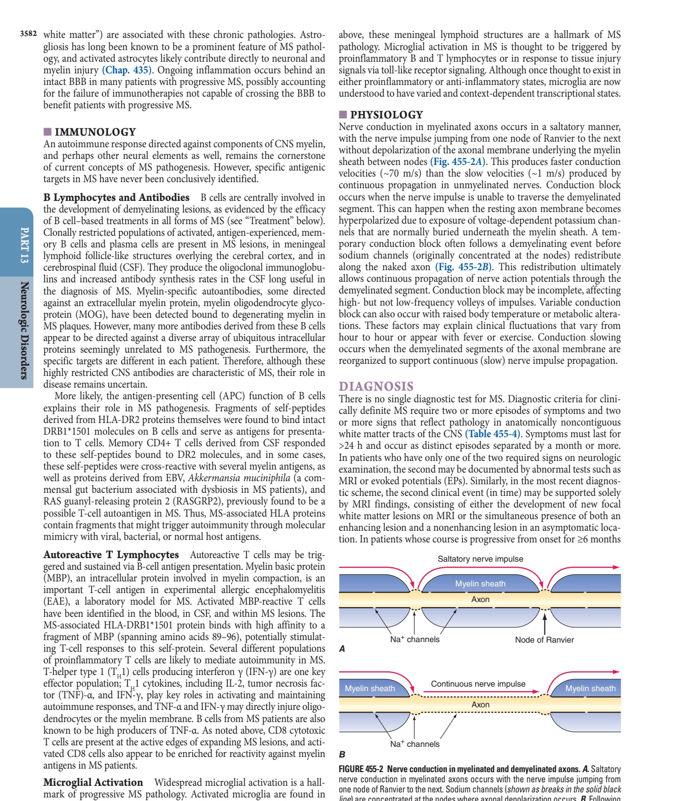
- Nerve conduction in myelinated axons occurs in a saltatory manner, with the nerve impulse jumping from one node of Ranvier to the next without depolarization of the axonal membrane underlying the myelin sheath between nodes.
- This produces faster conduction velocities (~70 m/s) than the slow velocities (~1 m/s) produced by continuous propagation in unmyelinated nerves.
- Conduction block occurs when the nerve impulse is unable to traverse the demyelinated segment.
- This can happen when the resting axon membrane becomes hyperpolarized due to exposure of voltage-dependent potassium channels that are normally buried underneath the myelin sheath.
- A temporary conduction block often follows a demyelinating event before sodium channels (originally concentrated at the nodes) redistribute along the naked axon.
- This redistribution ultimately allows continuous propagation of nerve action potentials through the demyelinated segment.
- Conduction block may be incomplete, affecting high- but not low-frequency volleys of impulses.
- Variable conduction block can also occur with raised body temperature or metabolic alterations.
- These factors may explain clinical fluctuations that vary from hour to hour or appear with fever or exercise.
- Conduction slowing occurs when the demyelinated segments of the axonal membrane are reorganized to support continuous (slow) nerve impulse propagation.
4. CLINICAL FEATURES
- Onset is typically between 20 and 40 years (slightly later in men than in women), but the disease can present across the lifespan.
- Women are affected approximately three times more often than men.
- Early symptoms may be severe or seem so trivial that a patient may not seek medical attention for months or years.
- On occasion, MS lesions are located exclusively in noneloquent regions of the nervous system, and in such instances, clinical manifestations can be largely or entirely absent.
- Autopsy series identified MS in some individuals (~0.1% of cases) who were seemingly asymptomatic during life, and magnetic resonance imaging (MRI) scans obtained for unrelated reasons also showed evidence of asymptomatic MS, an incidental finding termed a radiologically isolated syndrome (RIS).
- Specific symptoms of MS are varied and reflect the location and severity of lesions within the CNS.
- Neurologic examination often reveals unexpected findings in addition to the anticipated ones.
- For example, a patient may present with symptoms in one leg but signs in both.
4.1 Sensory Symptoms
- Sensory symptoms include both paresthesias (e.g., tingling, prickling sensations, 'pins and needles,' formications, or painful burning) and hypesthesia (e.g., reduced sensation, numbness, or a 'dead' feeling).
- Unpleasant sensations (e.g., feelings that body parts are swollen, wet, raw, or tightly wrapped) are also common.
- Sensory impairment of the trunk and legs below a horizontal line on the torso (a sensory level) indicates that the spinal cord is the site of the disturbance.
- It is often accompanied by a bandlike sensation of tightness around the torso.
- Pain is a common symptom of MS, experienced by >50% of patients.
- Pain can occur anywhere on the body and can change locations over time.
4.2 Visual Symptoms
- Optic neuritis (ON) presents as diminished visual acuity, dimness, or decreased color perception (desaturation) in the central field of vision.
- These symptoms can be mild or may progress to severe visual loss.
- Rarely, there is complete loss of light perception.
- Visual symptoms are generally monocular but may be bilateral.
- Periorbital pain (aggravated by eye movement) typically precedes or accompanies the visual loss.
- An afferent pupillary defect is usually present.
- Fundoscopic examination may be normal or reveal optic disc swelling (papillitis).
- Pallor of the optic disc (optic atrophy) commonly follows ON.
- Uveitis is uncommon and should raise the possibility of alternative diagnoses such as sarcoidosis or lymphoma.
4.3 Motor Symptoms
- Weakness of the limbs can manifest as loss of strength, speed, or blurring dexterity; as fatigue; or as a disturbance of gait.
- Exercise-induced weakness is a characteristic symptom of MS.
- The weakness is of the upper motor neuron type and is usually accompanied by other pyramidal signs such as spasticity, hyperreflexia, and extensor plantar responses.
- Occasionally, a tendon reflex may be lost (simulating a peripheral nerve lesion) if an MS lesion disrupts the afferent reflex (α-adrenergic innervation) fibers in the spinal cord.
- Facial weakness due to a lesion in the pons may resemble idiopathic Bell's palsy.
- Unlike Bell's palsy, facial weakness in MS is usually not associated with ipsilateral loss of taste sensation or retroauricular pain.
- Spasticity is commonly associated with spontaneous and movement-induced muscle spasms, especially in the legs.
- This can be accompanied by painful spasms interfering with ambulation, work, or self-care.
- Occasionally, spasticity provides support for the body weight during ambulation, and in these cases, treatment of spasticity may actually do more harm than good.
- Visual blurring in MS may result from ON or diplopia (double vision); if the symptom resolves when either eye is covered, the cause is diplopia.
- Diplopia may be caused by internuclear ophthalmoplegia (INO) or palsy of the sixth cranial nerve (rarely the third or fourth).
- An INO consists of impaired adduction of one eye due to a lesion in the ipsilateral medial longitudinal fasciculus.
- Prominent nystagmus is often observed in the abducting eye, along with a small skew deviation.
- A bilateral INO is particularly suggestive of MS.
- Other common gaze disturbances in MS include (1) a horizontal gaze palsy, (2) a 'one and a half' syndrome (horizontal gaze palsy plus an INO), and (3) acquired pendular nystagmus.
- Ataxia usually manifests as cerebellar tremors.
- Ataxia may also involve the head and trunk or the voice, producing a characteristic cerebellar dysarthria (scanning speech).
- Vertigo may appear suddenly from a brainstem lesion, superficially resembling acute labyrinthitis.
- Hearing loss may also occur in MS but is uncommon.
4.4 Ancillary Symptoms
- Paroxysmal symptoms are distinguished by their brief duration (10 s to 2 min), high frequency (5–40 episodes per day), lack of any alteration of consciousness or change in background electroencephalogram during episodes, and a self-limited course (generally lasting weeks to months).
- They may be precipitated by hyperventilation or movement.
- Manifestations can include Lhermitte's symptom; tonic contractions of a limb, face, or trunk (tonic seizures); paroxysmal dysarthria and ataxia; paroxysmal sensory disturbances; and several other less well-characterized syndromes.
- Paroxysmal symptoms probably result from spontaneous discharges arising at the edges of demyelinated plaques and spreading to adjacent white matter tracts.
- Lhermitte's symptom is an electric shock–like sensation (typically induced by flexion or other movements of the neck) that radiates down the back into the legs.
- Rarely, it radiates into the arms.
- It is generally self-limited but may persist for years.
- Lhermitte's symptom can also occur with other disorders of the cervical spinal cord (e.g., cervical spondylosis).
- Trigeminal neuralgia, hemifacial spasm, and glossopharyngeal neuralgia can occur when the demyelinating lesion involves the root entry (or exit) zone of the fifth, seventh, and ninth cranial nerve, respectively.
- Trigeminal neuralgia (tic douloureux) is a very brief lancinating facial pain often triggered by an afferent input from the face or teeth.
- Most cases of trigeminal neuralgia are not MS related; however, atypical features such as onset before age 50 years, bilateral symptoms, objective sensory loss, or nonparoxysmal pain should raise the possibility that MS could be responsible.
- Facial myokymia consists of either persistent rapid flickering contractions of the facial musculature (especially the lower portion of the orbicularis oculus) or a contraction that slowly spreads across the face.
- It results from lesions of the corticobulbar tracts or brainstem of the facial nerve.
- Heat sensitivity refers to neurologic symptoms produced by an elevation of the body's core temperature.
- For example, unilateral visual loss may occur during a hot shower or with physical exercise (Uhthoff's symptom).
- It is also common for MS symptoms to worsen transiently, sometimes dramatically, during febrile illnesses.
- Such heat-related symptoms probably result from transient conduction block.
- Bladder dysfunction is ultimately present in most MS patients.
- During normal reflex voiding, relaxation of the bladder sphincter is coordinated with contraction of the detrusor muscle in the bladder wall (muscarinic cholinergic innervation).
- Detrusor hyperreflexia, due to impairment of suprasegmental inhibition, causes urinary frequency, urgency, nocturia, and uncontrolled bladder emptying.
- Detrusor sphincter dyssynergia, due to loss of synchronization between detrusor and sphincter muscles, causes difficulty in initiating and/or stopping the urinary stream, producing hesitancy, urinary retention, overflow incontinence, and recurrent infection.
- Constipation occurs in some patients, especially with advanced disease.
- Fecal urgency or bowel incontinence is less common than urinary symptoms but can be socially debilitating.
- Sexual dysfunction may manifest as decreased libido, impaired genital sensation, impotence in men, and diminished vaginal lubrication or adductor spasms in women.
- Cognitive dysfunction is often mild when present, but can include memory loss; impaired attention; difficulties in executive functioning, memory, and problem solving; slowed information processing; and problems shifting between cognitive tasks.
- Euphoria (elevated mood) or emotional lability (pseudobulbar palsy) was once thought to be characteristic of MS but is actually relatively uncommon.
- Cognitive dysfunction sufficient to impair activities of daily living is rare.
- Depression, experienced by approximately half of patients, can be reactive, endogenous, or part of the illness itself and can contribute to fatigue.
- Fatigue is experienced by most MS patients and is the most common reason for work-related disability in MS.
- Fatigue can be exacerbated by elevated temperatures, depression, expending exceptional effort to accomplish basic activities of daily living, or sleep disturbances (e.g., from frequent nocturnal awakenings to urinate).
5. DIFFERENTIAL DIAGNOSIS
- There is no single diagnostic test for MS.
- Diagnostic criteria for clinically definite MS require two or more episodes of symptoms and two or more signs that reflect pathology in anatomically noncontiguous white matter tracts of the CNS.
- Symptoms must last for >24 h and occur as distinct episodes separated by a month or more.
- In patients who have only one of the two required signs on neurologic examination, the second may be documented by abnormal tests such as MRI or evoked potentials (EPs).
- Similarly, in the most recent diagnostic scheme, the second clinical event (in time) may be supported solely by MRI findings, consisting of either the development of new focal white matter lesions on MRI or the simultaneous presence of both an enhancing lesion and a nonenhancing lesion in an asymptomatic location.
- Uveitis is uncommon and should raise the possibility of alternative diagnoses such as sarcoidosis or lymphoma.
- Trigeminal neuralgia (tic douloureux) is a very brief lancinating facial pain often triggered by an afferent input from the face or teeth.
- Most cases of trigeminal neuralgia are not MS related; however, atypical features such as onset before age 50 years, bilateral symptoms, objective sensory loss, or nonparoxysmal pain should raise the possibility that MS could be responsible.
- Lhermitte's symptom can also occur with other disorders of the cervical spinal cord (e.g., cervical spondylosis).
6. INVESTIGATIONS & DIAGNOSIS
- There is no single diagnostic test for MS.
- Diagnostic criteria for clinically definite MS require two or more episodes of symptoms and two or more signs that reflect pathology in anatomically noncontiguous white matter tracts of the CNS.
- Symptoms must last for >24 h and occur as distinct episodes separated by a month or more.
- In patients who have only one of the two required signs on neurologic examination, the second may be documented by abnormal tests such as MRI or evoked potentials (EPs).
- Similarly, in the most recent diagnostic scheme, the second clinical event (in time) may be supported solely by MRI findings, consisting of either the development of new focal white matter lesions on MRI or the simultaneous presence of both an enhancing lesion and a nonenhancing lesion in an asymptomatic location.
- Myelin-specific autoantibodies, some directed against an extracellular myelin protein, myelin oligodendrocyte glycoprotein (MOG), have been detected bound to degenerating myelin in MS plaques.
- However, many more antibodies derived from these B cells appear to be directed against a diverse array of ubiquitous intracellular proteins seemingly unrelated to MS pathogenesis.
- Furthermore, the specific targets are different in each patient.
- Therefore, although these highly restricted CNS antibodies are characteristic of MS, their role in disease remains uncertain.
- More likely, the antigen-presenting cell (APC) function of B cells explains their role in MS pathogenesis.
- Fragments of self-peptides derived from HLA-DR2 proteins themselves were found to bind intact DRB1*1501 molecules on B cells and serve as antigens for presentation to T cells.
- Memory CD4+ T cells derived from CSF responded to these self-peptides bound to DR2 molecules, and in some cases, these self-peptides were cross-reactive with several myelin antigens, as well as proteins derived from EBV, Akkermansia muciniphila (a commensal gut bacterium associated with dysbiosis in MS patients), and RAS guanyl-releasing protein 2 (RASGRP2), previously found to be a possible T-cell autoantigen in MS.
- Thus, MS-associated HLA proteins contain fragments that might trigger autoimmunity through molecular mimicry with viral, bacterial, or norm
6.1 Diagnostic Criteria
- Diagnostic criteria for clinically definite MS require two or more episodes of symptoms and two or more signs that reflect pathology in anatomically noncontiguous white matter tracts of the CNS.
- Symptoms must last for >24 h and occur as distinct episodes separated by a month or more.
- In patients who have only one of the two required signs on neurologic examination, the second may be documented by abnormal tests such as MRI or evoked potentials (EPs).
- Similarly, in the most recent diagnostic scheme, the second clinical event (in time) may be supported solely by MRI findings, consisting of either the development of new focal white matter lesions on MRI or the simultaneous presence of both an enhancing lesion and a nonenhancing lesion in an asymptomatic location.
6.2 Scoring Systems
- Expanded Disability Status Scale (EDSS):
- 0.0 = Normal neurologic examination (all grade 0 in functional status [FS]).
- 1.0 = No disability, minimal signs in one FS (i.e., grade 1).
- 1.5 = No disability, minimal signs in more than one FS (more than one grade 1).
- 2.0 = Minimal disability in one FS (one FS grade 2, others 0 or 1).
- 2.5 = Minimal disability in two FS (two FS grade 2, others 0 or 1).
- 3.0 = Moderate disability in one FS (one FS grade 3, others 0 or 1) or mild disability in three or four FS (three/four FS grade 2, others 0 or 1) although fully ambulatory.
- 3.5 = Fully ambulatory but with moderate disability in one FS (one grade 3) and one or two FS grade 2; or two FS grade 3; or five FS grade 2 (others 0 or 1).
- 4.0 = Ambulatory without aid or rest for ~500 m.
- 4.5 = Ambulatory without aid or rest for ~300 m.
- 5.0 = Ambulatory without aid or rest for ~200 m.
- 5.5 = Ambulatory without aid or rest for ~100 m.
- 6.0 = Unilateral assistance required to walk about 100 m with or without resting.
- 6.5 = Constant bilateral assistance required to walk about 20 m without resting.
- 7.0 = Unable to walk beyond about 5 m even with aid; essentially restricted to wheelchair; wheels self and transfers alone.
- 7.5 = Unable to take more than a few steps; restricted to wheelchair; may need aid to transfer.
- 8.0 = Essentially restricted to bed or chair or perambulated in wheelchair, but out of bed most of day; retains many self-care functions; generally has effective use of arms.
- 8.5 = Essentially restricted to bed much of the day; has some effective use of arm(s); retains some self-care functions.
- 9.0 = Helpless bed patient; can communicate and eat.
- 9.5 = Totally helpless bed patient; unable to communicate or eat.
- 10.0 = Death due to MS.
- Functional Status (FS) Score:
- A. Pyramidal functions:
- 0 = Normal.
- 1 = Abnormal signs without disability.
- 2 = Minimal disability.
- 3 = Mild or moderate paraparesis or hemiparesis, or severe monoparesis.
- 4 = Marked paraparesis or hemiparesis, moderate quadriparesis, or monoplegia.
- 5 = Paraplegia, hemiplegia, or marked quadriparesis.
- 6 = Quadriplegia.
- B. Cerebellar functions:
- 0 = Normal.
- 1 = Abnormal signs without disability.
- 2 = Mild ataxia.
- 3 = Moderate truncal or limb ataxia.
- 4 = Severe ataxia all limbs.
- 5 = Unable to perform coordinated movements due to ataxia.
- C. Brainstem functions:
- 0 = Normal.
- 1 = Signs only.
- 2 = Moderate nystagmus or other mild disability.
- 3 = Severe nystagmus, marked extraocular weakness, or moderate disability of other cranial nerves.
- 4 = Marked dysarthria or other marked disability.
- 5 = Inability to swallow or speak.
- D. Sensory functions:
- 0 = Normal.
- 1 = Vibration or figure-writing decrease only, in 1 or 2 limbs.
- 2 = Mild decrease in touch or pain or position sense, and/or moderate decrease in vibration in 1 or 2 limbs, or vibratory decrease alone in 3 or 4 limbs.
- 3 = Moderate decrease in touch or pain or position sense, and/or essentially lost vibration in 1 or 2 limbs, or mild decrease in touch or pain, and/or moderate decrease in all proprioceptive tests in 3 or 4 limbs.
- 4 = Marked decrease in touch or pain or loss of proprioception, alone or combined, in 1 or 2 limbs or moderate decrease in touch or pain and/or severe proprioceptive decrease in >2 limbs.
- 5 = Loss (essentially) of sensation in 1 or 2 limbs or moderate decrease in touch or pain and/or loss of proprioception for most of the body below the head.
- 6 = Sensation essentially lost below the head.
- E. Bowel and bladder functions:
- 0 = Normal.
- 1 = Mild urinary hesitancy, urgency, or retention.
- 2 = Moderate hesitancy, urgency, retention of bowel or bladder, or rare urinary incontinence.
- 3 = Frequent urinary incontinence.
- 4 = In need of almost constant catheterization.
- 5 = Loss of bladder function.
- 6 = Loss of bowel and bladder function.
- F. Visual (or optic) functions:
- 0 = Normal.
- 1 = Scotoma with visual acuity (corrected) better than 20/30.
- 2 = Worse eye with scotoma with maximal visual acuity (corrected) of 20/30 to 20/59.
- 3 = Worse eye with large scotoma, or moderate decrease in fields, but with maximal visual acuity (corrected) of 20/60 to 20/99.
- 4 = Worse eye with marked decrease of fields and maximal acuity (corrected) of 20/100 to 20/200; grade 3 plus maximal acuity of better eye of 20/60 or less.
- 5 = Worse eye with maximal visual acuity (corrected) <20/200; grade 4 plus maximal acuity of better eye of ≤20/60.
- 6 = Grade 5 plus maximal visual acuity of better eye of ≤20/60.
- G. Cerebral (or mental) functions:
- 0 = Normal.
- 1 = Mood alteration only (does not affect EDSS score).
- 2 = Mild decrease in mentation.
- 3 = Moderate decrease in mentation.
- 4 = Marked decrease in mentation.
- 5 = Chronic brain syndrome—severe or incompetent.
7. MANAGEMENT & TREATMENT
- The past decade has seen tremendous progress in understanding basic disease mechanisms underlying MS and in developing highly effective therapies especially for the relapsing form of the disease.
- These advances have dramatically improved the long-term outcome for patients.
- B cell–based treatments in all forms of MS.
- Highly effective therapies, attacks are abolished in most patients, but insidious progression independent of relapse activity, termed 'silent progression,' is now evident during the relapsing phase.
- Immunotherapies not capable of crossing the BBB to benefit patients with progressive MS.
7.1 Therapeutic Goals
- Promoting remyelination to protect axons remains an important therapeutic goal.
- Ongoing inflammation occurs behind an intact BBB in many patients with progressive MS, possibly accounting for the failure of immunotherapies not capable of crossing the BBB to benefit patients with progressive MS.
7.2 Treatment Classes
- B cell–based treatments in all forms of MS.
- Highly effective therapies for MS.
- Immunotherapies not capable of crossing the BBB to benefit patients with progressive MS.
8. PROGNOSIS & COMPLICATIONS
- Disability in MS is thought to accumulate as either a consequence of limited recovery following an acute relapse, a process also known as relapse associated worsening (RAW), or from presumed underlying neurodegeneration in the absence of clinical relapse, a process termed progression independent of relapsing activity (PIRA).
- Although RAW was once thought to be the primary driver of disability accumulation in RRMS, it is now clear that PIRA is the cause of disability accumulation in RRMS, SPMS, and PPMS.
- That PIRA events can occur 'silently,' meaning so insidiously that neither the patient nor the provider recognizes their occurrence at the time of gradual worsening, raises the important question as to whether there is a meaningful distinction between RRMS and SPMS.
- If any confirmed PIRA event is considered to be indicative of SPMS, then the SPMS onset begins much earlier in the disease course when patients still experience relapses but have only accumulated relative minor disability.
- In the pretreatment era, the risk of developing SPMS was ~3% each year, meaning that the great majority of RRMS would ultimately evolve into SPMS.
- However, more recent case series have indicated a much lower rate of evolution to SPMS, estimated at <1% each year, likely due to widespread use of increasingly effective therapies for MS.
- Disability accumulation is driven by 'silent progression' (PIRA) independent of relapse activity in the treatment era.
9. SPECIAL CONSIDERATIONS
- In the United States, there is a slightly higher prevalence in white compared with black individuals, with lower estimates in Hispanics, followed by Asians.
- The prevalence of MS also increased steadily in several regions around the world over the past half-century, presumably reflecting the impact of some environmental shift, improved diagnosis, and/or a longer lifespan.
- Moreover, this increase appears to have occurred to a greater degree in women than men and in nonwhite populations.
10. KEY PEARLS & CLINICAL TRAPS
- Women are affected approximately three times more often than men.
- Onset is typically between 20 and 40 years (slightly later in men than in women).
- Heat sensitivity (Uhthoff's symptom) and Lhermitte's symptom are characteristic clinical features.
- Pain is a common symptom of MS, experienced by >50% of patients.
- A bilateral INO is particularly suggestive of MS.
- Cognitive dysfunction sufficient to impair activities of daily living is rare.
- Euphoria (elevated mood) or emotional lability (pseudobulbar palsy) was once thought to be characteristic of MS but is actually relatively uncommon.
- Fatigue is experienced by most MS patients and is the most common reason for work-related disability in MS.
- Autopsy series identified MS in some individuals (~0.1% of cases) who were seemingly asymptomatic during life.
- EBV infection is a near-universal prerequisite for development of MS.
- HLA-DRB1*1501 is the strongest genetic susceptibility signal, accounting for ~10% of disease risk.
- In the treatment era, attacks are abolished in most patients, but insidious progression independent of relapse activity, termed 'silent progression,' is now evident during the relapsing phase.
Figures & Illustrations
Reproduced from Harrison's 22nd Edition.
Figure 1
Caption: FIGURE 455-2 Nerve conduction in myelinated and demyelinated axons. A. Saltatory nerve conduction in myelinated axons occurs with the nerve impulse jumping from one node of Ranvier to the next. Sodium channels (shown as breaks in the solid black in line) are concentrated at the nodes where axonal depolarization occurs. B. Following demyelination, additional sodium channels are redistributed along the axon itself, thereby allowing continuous propagation of the nerve action potential despite the absence of myelin. — Figure 455-1: The clinical course of multiple sclerosis (MS) in the current treatment era. The top half illustrates the traditional view of the natural history of relapse-onset MS in the pretreatment era, showing disability accumulation from incomplete recovery. The bottom half shows the new natural history in the current treatment era, where attacks are abolished but insidious progression independent of relapse activity ('silent progression') is evident.
Figure 2
Caption: FIGURE 455-1 The clinical course of multiple sclerosis (MS) in the current treatment era. The top half of the figure illustrates the traditional view of the natural history of relapse-onset MS in the pretreatment era. During the relapsing phase, disability accumulation was thought to result from incomplete recovery from relapses, until relapse- independent disability, designated SPMS, supervened. In the bottom half of the figure, the “new” natural history of MS in the current treatment era is shown. With use of highly effective therapies, attacks are abolished in most patients, but insidious progression independent of relapse activity, termed “silent progression,” is now evident during the relapsing phase. CIS, clinically isolated syndrome; EDSS, Extended Disability Status Score; MRI, magnetic resonance imaging; RIS, radiologically isolated syndrome; SPMS, secondary progressive multiple sclerosis. — Figure 455-2A: Saltatory conduction in myelinated axons. The nerve impulse jumps from one node of Ranvier to the next, producing faster conduction velocities (~70 m/s) compared to unmyelinated nerves.
Figure 3
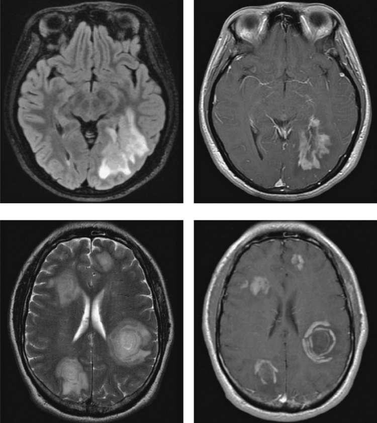
Caption: FIGURE 455-4 Magnetic resonance imaging findings in variants of MS. A and B. Acute (FLAIR) image of a large solitary right parieto-occipital white matter lesion is shown, with image obtained after the intravenous administration of gadolinium diethylene triamine disruption consistent with acute inflammation. C and D. Balo’s concentric sclerosis. In signal in the supratentorial white matter bilaterally; some lesions reveal concentric images after gadolinium demonstrate abnormal enhancement of all lesions with some activity, but more recent studies indicate that these produce little if any additional risk. — Figure 455-2B: Conduction block in demyelinated segments. When the resting axon membrane becomes hyperpolarized due to exposure of voltage-dependent potassium channels, conduction block occurs. Sodium channels redistribute along the naked axon to allow continuous propagation.
Figure 4
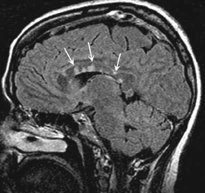
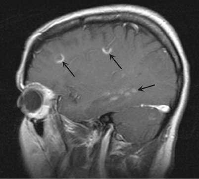
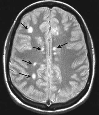
Caption: FIGURE 455-3 Magnetic resonance imaging findings in multiple sclerosis (MS). A. Axial abnormalities in white matter, typical for MS. B. Sagittal T2-weighted fluid-attenuated has been suppressed. CSF appears dark, whereas areas of brain edema or demyelination the anterior corpus callosum are frequent in MS and rare in vascular disease. C. Sagittal high-signal-intensity lesion in the midthoracic spinal cord. D. Sagittal T1-weighted pentaacetic acid (DTPA) reveals focal areas of blood-brain barrier disruption, identified — Figure 455-3: MRI findings in multiple sclerosis. Demonstrates typical periventricular white matter lesions and demyelinating plaques characteristic of MS.
Figure 5
Caption: FIGURE 455-3 Magnetic resonance imaging findings in multiple sclerosis (MS). A. Axial abnormalities in white matter, typical for MS. B. Sagittal T2-weighted fluid-attenuated has been suppressed. CSF appears dark, whereas areas of brain edema or demyelination the anterior corpus callosum are frequent in MS and rare in vascular disease. C. Sagittal high-signal-intensity lesion in the midthoracic spinal cord. D. Sagittal T1-weighted pentaacetic acid (DTPA) reveals focal areas of blood-brain barrier disruption, identified — Figure 455-4: Optic neuritis MRI findings. Shows optic nerve enhancement and signal abnormalities consistent with inflammation and demyelination.
Figure 6
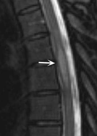
Caption: FIGURE 455-3 Magnetic resonance imaging findings in multiple sclerosis (MS). A. Axial abnormalities in white matter, typical for MS. B. Sagittal T2-weighted fluid-attenuated has been suppressed. CSF appears dark, whereas areas of brain edema or demyelination the anterior corpus callosum are frequent in MS and rare in vascular disease. C. Sagittal high-signal-intensity lesion in the midthoracic spinal cord. D. Sagittal T1-weighted pentaacetic acid (DTPA) reveals focal areas of blood-brain barrier disruption, identified — Figure 455-5: Spinal cord MRI in MS. Illustrates intramedullary lesions in the cervical or thoracic spinal cord causing sensory or motor deficits.
Figure 7
Caption: FIGURE 455-3 Magnetic resonance imaging findings in multiple sclerosis (MS). A. Axial abnormalities in white matter, typical for MS. B. Sagittal T2-weighted fluid-attenuated has been suppressed. CSF appears dark, whereas areas of brain edema or demyelination the anterior corpus callosum are frequent in MS and rare in vascular disease. C. Sagittal high-signal-intensity lesion in the midthoracic spinal cord. D. Sagittal T1-weighted pentaacetic acid (DTPA) reveals focal areas of blood-brain barrier disruption, identified — Figure 455-6: Cortical lesions in MS. Shows subpial cortical demyelination and ectopic lymphoid follicles associated with neurodegeneration.
Generated from Harrison's Principles of Internal Medicine, 22nd Edition.