Mitral Stenosis and Mitral Regurgitation
Chapter 274 & 275 | Part 6: Disorders of the Cardiovascular System · Part 6 – Cardiovascular Disorders
Detailed clinical reference synthesised from Harrison's Principles of Internal Medicine, 22nd Edition
🔑 Key Clinical Points
- Rheumatic fever is the leading cause of mitral stenosis (MS); incidence has declined in temperate climates but remains high in low-income countries.
- Severe MS is defined by a mitral valve area (MVA) <1.5 cm2; critical stenosis is <1.0 cm2.
- The opening snap (OS) follows aortic valve closure (A) by 0.05–0.12 s; the interval varies inversely with MS severity.
- Percutaneous mitral balloon commissurotomy (PMBC) is preferred for symptomatic patients with pliable valves and no significant MR or thrombus.
- Vitamin K antagonists (INR 2–3) are recommended for MS patients with AF or history of thromboembolism; DOACs are not recommended for rheumatic MS.
- Chronic severe MR is defined by regurgitant volume ≥60 mL/beat, RF ≥50%, and effective regurgitant orifice area ≥0.40 cm2.
- In acute severe MR, the arterial pressure may be reduced with a narrow pulse pressure, and signs of pulmonary congestion are prominent.
- Mitral annular calcification may include elements of both primary and secondary MR (mixed) as the disease process may encroach on the leaflets.
- Systemic embolization in MS occurs more frequently in patients with AF, age >65 years, and reduced CO.
- Surgical mitral valve replacement (MVR) is necessary in patients with MS and significant associated MR or severe distortion by previous manipulation.
📑 Table of Contents
- 1. DEFINITION & OVERVIEW
- 1.1 Mitral Stenosis (MS)
- 1.2 Mitral Regurgitation (MR)
- 2. EPIDEMIOLOGY
- 2.1 Mitral Stenosis Epidemiology
- 3. ETIOLOGY & PATHOPHYSIOLOGY
- 3.1 Mitral Stenosis Pathophysiology
- 3.2 Mitral Regurgitation Pathophysiology
- 4. CLINICAL FEATURES
- 4.1 Mitral Stenosis Physical Findings
- 4.2 Mitral Regurgitation Physical Findings
- 5. DIFFERENTIAL DIAGNOSIS
- 5.1 Mitral Stenosis Differential Diagnosis
- 5.2 Mitral Regurgitation Differential Diagnosis
- 6. INVESTIGATIONS & DIAGNOSIS
- 6.1 Mitral Stenosis Investigations
- 6.2 Mitral Regurgitation Investigations
- 7. MANAGEMENT & TREATMENT
- 7.1 Mitral Stenosis Management
- 7.2 Mitral Regurgitation Management
- 8. PROGNOSIS & COMPLICATIONS
- 8.1 Mitral Stenosis Complications
- 8.2 Mitral Regurgitation Complications
- 9. SPECIAL CONSIDERATIONS
- 9.1 Mitral Stenosis Special Considerations
- 10. KEY PEARLS & CLINICAL TRAPS
- 10.1 Mitral Stenosis Pearls
- 10.2 Mitral Regurgitation Pearls
- Flowcharts & Algorithms
- Figures & Illustrations
📋 Figures in This Chapter
| # | Type | Description |
|---|---|---|
| 1 | 🔀 Flowchart | Management of rheumatic mitral stenosis |
| 2 | 🔀 Flowchart | Mitral regurgitation |
| 3 | 🔀 Flowchart | Management of secondary mitral regurgitation |
| 1 | 🖼 Figure | Clip devices used to grasp the free edges of the anterior and... |
| 2 | 🖼 Figure | MVR is now routinely performed with preservation of the chordal FIGURE 274-3... |
| 3 | 🖼 Figure | Continuous wave Doppler interrogation of transmitral valve velocities in a patient with... |
| 4 | 🖼 Figure | Figure / Illustration |
1. DEFINITION & OVERVIEW
Mitral stenosis (MS) is a valvular heart disease characterized by obstruction of left ventricular inflow due to narrowing of the mitral valve orifice. Mitral regurgitation (MR) is the backflow of blood from the left ventricle into the left atrium during systole. Both conditions are major causes of morbidity and mortality worldwide.
1.1 Mitral Stenosis (MS)
MS is primarily caused by rheumatic fever. Chronic inflammation leads to diffuse thickening of the valve leaflets with formation of fibrous tissue often with calcific deposits. The mitral commissures fuse, the chordae tendineae fuse and shorten, the valvular cusps become rigid, and the pathologic process eventually leads to narrowing at the apex of the funnel-shaped (fish-mouth) valve. Although the initial insult is rheumatic, later changes may be exacerbated by inflammation, fibrosis, and trauma due to altered flow patterns. Calcification of the stenotic mitral valve immobilizes the leaflets and narrows the orifice further.
Table 1 — Table 274-1 Major Causes of Mitral Stenosis
| Etiologies |
|---|
| Rheumatic fever |
| Congenital (parachute valve, cor triatriatum) |
| Severe mitral annular calcification with leaflet involvement |
| SLE, RA |
| Myxoma |
| IE with large vegetations |
1.2 Mitral Regurgitation (MR)
MR may result from an abnormality or disease process that affects any one or more of the five functional components of the mitral valve apparatus (leaflets, annulus, chordae tendineae, papillary muscles, and subjacent myocardium). Distinction should be drawn between primary (degenerative) MR, in which the leaflets and/or chordae tendineae are primarily responsible for abnormal valve function, and secondary (functional) MR, in which the leaflets and chordae tendineae are usually normal but the regurgitation is caused by left ventricular (LV) and/or left atrial remodeling, annular dilation, papillary muscle displacement, dysynchrony, leaflet tethering, or their combination.
Table 2 — Table 275-1 Major Causes of Mitral Regurgitation (MR)
| Etiologies |
|---|
| Acute: IE |
| Papillary muscle rupture (post-MI) |
| Chordal rupture/leaflet flail (MVP, IE) |
| Blunt trauma |
| Chronic: Primary (affecting leaflets, chordae) |
| Myxomatous (MVP, Barlow's, forme fruste) |
| Rheumatic fever |
| IE (healed) |
| Congenital (cleft, AV canal) |
| Radiation |
| Secondary (leaflets, chordae are 'innocent bystanders'): |
| Ischemic cardiomyopathy |
| Dilated cardiomyopathy |
| HOCM (with SAM) |
| AF with LA enlargement and annular dilation (atrial functional MR) |
| Mitral annular calcificationa |
2. EPIDEMIOLOGY
With reductions in the incidence of acute rheumatic fever, particularly in temperate climates and middle- to high-income countries, the incidence of MS has declined considerably over the past several decades. However, it remains a major problem in low-income countries, especially in sub-Saharan Africa, India, Southeast Asia, and Oceania. Pure or predominant MS occurs in ~40% of all patients with rheumatic heart disease and a history of rheumatic fever. In other patients with rheumatic heart disease, lesser degrees of MS may accompany mitral regurgitation (MR) and aortic valve disease.
2.1 Mitral Stenosis Epidemiology
In temperate climates, the latent period between the initial attack of rheumatic carditis (in the increasingly rare circumstances in which a history of one can be elicited) and the development of symptoms due to MS is generally about two decades; most patients begin to experience disability in the fourth decade of life. Studies carried out before the development of surgical mitral valvotomy revealed that once a patient with MS became seriously symptomatic, the disease progressed inexorably to death within 2–5 years.
3. ETIOLOGY & PATHOPHYSIOLOGY
In rheumatic MS, chronic inflammation leads to diffuse thickening of the valve leaflets with formation of fibrous tissue often with calcific deposits. The mitral commissures fuse, the chordae tendineae fuse and shorten, the valvular cusps become rigid, and the pathologic process eventually leads to narrowing at the apex of the funnel-shaped (fish-mouth) valve. Although the initial insult to the mitral valve is rheumatic, later changes may be exacerbated by inflammation, fibrosis, and trauma due to altered flow patterns. Calcification of the stenotic mitral valve immobilizes the leaflets and narrows the orifice further. Thrombus formation and arterial embolization may arise from the calcific valve itself, but in patients with atrial fibrillation (AF), thrombi arise more frequently from the dilated left atrium (LA), particularly from within the LA appendage.
3.1 Mitral Stenosis Pathophysiology
In normal adults, the area of the mitral valve orifice is 4–6 cm2. In the presence of significant obstruction, i.e., when the orifice area is reduced to <~2 cm2, blood can flow from the LA to the left ventricle (LV) only if propelled by an abnormally elevated left atrioventricular pressure gradient, the hemodynamic hallmark of MS. When the mitral valve opening is reduced to <1.5 cm2, referred to as severe MS, an LA pressure of ~25 mmHg is required to maintain a normal cardiac output (CO). The elevated pulmonary venous and pulmonary arterial (PA) wedge pressures reduce pulmonary compliance, contributing to exertional dyspnea. The first bouts of dyspnea are usually precipitated by sudden changes in the heart rate, volume status, or CO, as, for example, with severe exertion, excitement, fever, severe anemia, paroxysmal AF and other tachycardias, sexual intercourse, pregnancy, and thyrotoxicosis. As MS progresses, lesser degrees of stress precipitate dyspnea, the patient becomes limited in daily activities, and orthopnea and paroxysmal nocturnal dyspnea develop. The development of persistent AF often marks a turning point in the patient's course and is generally associated with acceleration of the rate at which symptoms progress. Hemoptysis (Chap. 41) results from rupture of pulmonary-bronchial venous connections secondary to pulmonary venous hypertension. It occurs most frequently in patients who have elevated LA pressures without markedly elevated pulmonary vascular resistances and is rarely fatal. Recurrent pulmonary emboli (Chap. 290), sometimes an important cause of morbidity and mortality late in the course of MS and often arise from right atrial mural thrombus. Pulmonary infections, i.e., bronchitis, bronchopneumonia, and lobar pneumonia, commonly complicate untreated MS, especially during the winter months.
3.2 Mitral Regurgitation Pathophysiology
The resistance to LV emptying (LV afterload) is reduced in patients with MR. As a consequence, the LV is decompressed into the LA during ejection, and with the reduction in LV size during systole, there is a rapid decline in LV tension. The initial compensation to MR is more complete LV emptying. However, LV volume increases progressively with time as the severity of the regurgitation increases and as LV contractile function deteriorates. This increase in LV volume is often accompanied by a reduced forward cardiac output (CO). LV compliance is often increased, and thus, LV diastolic pressure does not increase until late in the course. The regurgitant volume varies directly with the LV systolic pressure and the size of the regurgitant orifice; the latter, in turn, is influenced by the extent of LV and mitral annular dilation, as well as by leaflet morphology. Because ejection fraction (EF) rises in severe MR in the presence of normal LV function, even a modest reduction in this parameter (<60%) reflects significant contractile dysfunction. During early diastole, as the distended LA empties, there is a particularly rapid y descent in the absence of accompanying MS. A brief, early diastolic LA-LV pressure gradient (often generating a rapid filling sound [S] and mid-diastolic murmur masquerading as MS) may occur in patients with pure, severe MR as a result of the very rapid flow of blood across a normal-sized mitral orifice.
4. CLINICAL FEATURES
In patients with severe MS, there may be a malar flush with pinched and blue facies. In patients with sinus rhythm and severe pulmonary hypertension or associated TS, the jugular venous pulse reveals prominent a waves due to vigorous right atrial systole. The systemic arterial pressure is usually normal or slightly low. A parasternal lift signifies an enlarged RV. A diastolic thrill may very rarely be present at the cardiac apex, with the patient in the left lateral recumbent position.
4.1 Mitral Stenosis Physical Findings
Auscultation: The first heart sound (S1) is usually accentuated in the early stages of the disease and slightly delayed. The pulmonic component of the second heart sound (P2) also is often accentuated with elevated PAPs, and the two components of the second heart sound (S2) are closely split. The opening snap (OS) of the mitral valve is most readily audible in expiration at, or just medial to, the cardiac apex. This sound generally follows the sound of aortic valve closure (A2) by 0.05–0.12 s. The time interval between A2 and OS varies inversely with the severity of the MS. The OS is followed by a low-pitched, rumbling, diastolic murmur, heard best at the apex with the patient in the left lateral recumbent position (see Fig. 246-5); it is accentuated by mild exercise (e.g., a few rapid sit-ups) carried out just before auscultation. In general, the duration of this murmur correlates with the severity of the stenosis in patients with preserved CO. In patients with sinus rhythm, the murmur often reappears or becomes louder during atrial systole (presystolic accentuation). Soft, grade I or II/VI systolic murmurs may be heard at or medial to the apex and may signify mixed mitral valve disease with regurgitation. Hepatomegaly, ankle edema, ascites, and pleural effusion, particularly in the right pleural cavity, may occur in patients with MS and RV failure. Associated Lesions: With severe pulmonary hypertension, a pansystolic murmur produced by functional TR may be audible along the left sternal border. This murmur is usually louder during inspiration and diminishes during forced expiration (Carvallo's sign). When the CO is markedly reduced in MS, the typical auscultatory findings, including the diastolic rumbling murmur, may not be detectable (silent MS), but they may reappear as compensation is restored. The Graham Steell murmur of PR, a high-pitched, diastolic, decrescendo blowing murmur along the left sternal border, results from dilation of the pulmonary valve ring and occurs in patients with mitral valve disease and severe pulmonary hypertension. This murmur may be indistinguishable from the more common murmur produced by aortic regurgitation (AR), although it may increase in intensity with inspiration and is accompanied by a loud and often palpable P2.
4.2 Mitral Regurgitation Physical Findings
In patients with chronic severe MR, the arterial pressure is usually normal, although the carotid arterial pulse may show a sharp, low-volume upstroke owing to the reduced forward CO. A systolic thrill is often palpable at the cardiac apex, the LV is hyperdynamic with a brisk systolic impulse and a palpable rapid-filling wave (S3), and the apex beat is often displaced laterally. Auscultation: S1 is generally absent, soft, or buried in the holosystolic murmur of chronic, severe MR. In patients with severe MR, the aortic valve may close prematurely (due to the reduced forward cardiac output), resulting in wide but physiologic splitting of S2. A low-pitched S3 occurring 0.12–0.17 s after the aortic valve closure sound, i.e., at the completion of the rapid-filling phase of the LV, is believed to be caused by the sudden tensing of the papillary muscles, chordae tendineae, and valve leaflets. It may be followed by a short, rumbling, mid-diastolic murmur, even in the absence of structural MS. In patients with ischemic or dilated cardiomyopathy, however, a third sound (S3) may also signify ventricular dysfunction. A fourth heart sound is often audible in patients with acute severe MR who are in sinus rhythm. A presystolic murmur is not ordinarily heard with isolated MR. A systolic murmur of at least grade III/VI intensity is the most characteristic auscultatory finding in chronic severe MR. It is usually holosystolic (see Fig. 246-5A), but as previously noted, it is decrescendo and ceases in mid-to-late systole in patients with acute severe MR. The systolic murmur of chronic MR is usually most prominent at the apex and radiates to the axilla. However, in patients with ruptured chordae tendineae or primary involvement of the posterior mitral leaflet with prolapse or flail, the regurgitant jet is eccentric, directed anteriorly, and strikes the LA wall adjacent to the aortic root. In this situation, the systolic murmur is transmitted to the base of the heart and, therefore, may be confused with the murmur of AS. The murmur associated with anterior leaflet prolapse or flail is directed to the axilla. In patients with ruptured chordae tendineae, the systolic murmur may have a cooing or seagull quality, whereas a flail leaflet may produce a murmur with a musical quality. The systolic murmur of chronic MR not due to MVP is intensified by isometric exercise (handgrip) but is reduced during the strain phase of the Valsalva maneuver because of the associated decrease in LV preload.
5. DIFFERENTIAL DIAGNOSIS
Like MS, significant MR may also be associated with a prominent diastolic murmur at the apex due to increased antegrade transmitral flow, but in patients with isolated MR, this diastolic murmur commences slightly later than in patients with MS, and there is often clear-cut evidence of LV enlargement. An OS and increased P2 are absent, and S1 is soft or absent. An apical pansystolic murmur of at least grade III/VI intensity as well as an S3 suggests significant MR. Similarly, the apical mid-diastolic murmur associated with severe AR (Austin Flint murmur) may be mistaken for MS but can be differentiated from it because it is not intensified in pre-systole and becomes softer with administration of amyl nitrite or other arterial vasodilators. TS, which occurs rarely in the absence of MS, may mask many of the clinical features of MS or be clinically silent; when present, the diastolic murmur of TS increases with inspiration and the y descent in the jugular venous pulse is delayed.
5.1 Mitral Stenosis Differential Diagnosis
Atrial septal defect (Chap. 280) may be mistaken for MS; in both conditions, there is often clinical, ECG, and chest x-ray evidence of RV enlargement and accentuation of pulmonary vascularity. However, the absence of LA enlargement and of Kerley B lines and the demonstration of fixed splitting of S2 with a grade II or III mid-systolic murmur at the mid to upper left sternal border all favor atrial septal defect over MS. Atrial septal defects with large left-to-right shunts may result in functional TS because of the enhanced diastolic flow. An incomplete right bundle branch block pattern on ECG is often present. Left atrial myxoma (Chap. 282) may obstruct LA emptying, causing dyspnea, a diastolic murmur, and hemodynamic changes resembling those of MS. However, patients with an LA myxoma often have features suggestive of a systemic disease, such as weight loss, fever, anemia, systemic emboli, and elevated serum IgG and interleukin 6 (IL-6) concentrations. The auscultatory findings may change markedly with body position. The diagnosis can be established by the demonstration of a characteristic echo-producing mass in the LA with TTE.
5.2 Mitral Regurgitation Differential Diagnosis
Austin Flint murmur: The apical mid-diastolic murmur associated with severe AR may be mistaken for MS but can be differentiated from it because it is not intensified in pre-systole and becomes softer with administration of amyl nitrite or other arterial vasodilators. TS: TS, which occurs rarely in the absence of MS, may mask many of the clinical features of MS or be clinically silent; when present, the diastolic murmur of TS increases with inspiration and the y descent in the jugular venous pulse is delayed.
6. INVESTIGATIONS & DIAGNOSIS
ECG: In MS and sinus rhythm, the P wave usually suggests LA enlargement (see Fig. 247-8). It may become tall and peaked in lead II and upright in lead V1 when severe pulmonary hypertension or TS complicates MS and right atrial (RA) enlargement develops. The QRS complex is usually normal. However, with severe pulmonary hypertension, right axis deviation and RV hypertrophy are often present. In patients with sinus rhythm and severe pulmonary hypertension or associated TS, the jugular venous pulse reveals prominent a waves due to vigorous right atrial systole. The systemic arterial pressure is usually normal or slightly low. A parasternal lift signifies an enlarged RV. A diastolic thrill may very rarely be present at the cardiac apex, with the patient in the left lateral recumbent position.
6.1 Mitral Stenosis Investigations
Echocardiogram (TTE): Transthoracic echocardiography (TTE) with color flow and spectral Doppler imaging provides critical information, including measurements of mitral inflow velocity during early (E wave) and late (A wave in patients in sinus rhythm) diastolic filling, estimates of the transvalvular peak and mean gradients and mitral orifice area (Fig. 274-1), the presence and severity of any associated MR, the extent of leaflet calcification and restriction, the degree of distortion of the subvalvular apparatus, and the anatomic suitability for percutaneous mitral balloon commissurotomy (PMBC; see below). In addition, TTE provides an assessment of LV and RV function, chamber sizes, an estimation of the PA systolic pressure based on the tricuspid regurgitant jet velocity, and an indication of the presence and severity of any associated valvular lesions, such as aortic stenosis (AS) and/or regurgitation. Transesophageal echocardiography (TEE) provides superior images and should be used when TTE is inadequate for guiding management decisions. TEE is especially indicated to exclude the presence of LA thrombus prior to PMBC. The performance of TTE with exercise to evaluate the mean mitral diastolic gradient, PAPs, and RV function can be very helpful in the evaluation of patients with MS when there is a discrepancy between the clinical findings and the resting hemodynamics. Chest X-Ray: The earliest changes are straightening of the upper left border of the cardiac silhouette, prominence of the main PAs, dilation of the upper lobe pulmonary veins, and posterior displacement of the esophagus by an enlarged LA. Kerley B lines are fine, dense, opaque, horizontal lines that are most prominent in the lower and mid-lung fields that result from distention of interlobular septae and lymphatics with edema when the resting mean LA pressure exceeds ~20 mmHg.
6.2 Mitral Regurgitation Investigations
Echocardiogram (TTE): Transthoracic echocardiography (TTE) is indicated to assess the mechanism of the MR and its hemodynamic severity. LV function can be assessed from LV end-diastolic and end-systolic volumes and EF. Observations can be made regarding leaflet structure and function, chordal integrity, LA and LV size, annular calcification, and regional and global LV systolic function. Doppler imaging should demonstrate the width or area of the color flow MR jet within the LA, the duration and intensity of the continuous wave Doppler signal, the pulmonary venous flow contour, the early peak mitral inflow velocity, and quantitative measures of regurgitant volume, RF, and effective regurgitant orifice area. In addition, the PA pressures (PAPs) can be estimated from the TR jet velocity. TTE is also indicated to follow the course of patients with chronic MR and to provide rapid assessment for any clinical change. Transesophageal echocardiography (TEE) provides greater anatomic detail than TTE (see Fig. 248-5). Chest X-Ray: The LA and LV are the dominant chambers in chronic MR. Late in the course of the disease, the LA may be massively enlarged and forms the right border of the cardiac silhouette. Patients with acute severe MR may have asymmetric pulmonary edema if the regurgitant jet is directed predominantly to the orifice of an upper lobe pulmonary vein.
7. MANAGEMENT & TREATMENT
Penicillin prophylaxis of group A β-hemolytic streptococcal infections (Chap. 371) for secondary prevention of rheumatic fever is important for at-risk patients with rheumatic MS. Recommendations for infective endocarditis prophylaxis are similar to those for other valve lesions and are restricted to patients at high risk for complications from infection, including patients with a history of endocarditis. In symptomatic patients, some improvement usually occurs with restriction of sodium intake and small doses of oral diuretics. Beta blockers, nondihydropyridine calcium channel blockers (e.g., verapamil or diltiazem), and digitalis glycosides are useful in slowing the ventricular rate of patients with AF. Vitamin K antagonist therapy (such as warfarin) targeted to an international normalized ratio (INR) of 2–3 should be administered indefinitely to patients with MS who have AF, a history of thromboembolism, or demonstrated LA thrombus. The routine use of a vitamin K antagonist in patients in sinus rhythm with LA enlargement (maximal dimension >5.5 cm) with or without spontaneous echo contrast is more controversial. In a randomized trial of patients with rheumatic MS and AF, there was a significantly higher incidence of death among patients treated with rivaroxaban than with vitamin K antagonist therapy. A vitamin K antagonist is recommended to reduce the risk for stroke or systemic embolism in at-risk patients with rheumatic MS.
7.1 Mitral Stenosis Management
Cardioversion: If AF is of relatively recent onset in a patient whose MS is not severe enough to warrant PMBC or surgical intervention, reversion to sinus rhythm pharmacologically or by means of electrical countershock is indicated. Usually, cardioversion should be undertaken after the patient has had at least 3 consecutive weeks of anticoagulant treatment to a therapeutic INR. If cardioversion is indicated more urgently, then intravenous heparin should be provided and TEE performed to exclude the presence of LA thrombus before the procedure. Conversion to sinus rhythm is rarely successful or sustained in patients with severe MS, particularly those in whom the LA is significantly enlarged or in whom AF has been present for >1 year, conditions that favor the development of an LA myopathy. Mitral Commissurotomy: Unless there is a contraindication, mitral commissurotomy is indicated in symptomatic (New York Heart Association [NYHA] functional class II–IV) patients with isolated severe MS, whose effective orifice (valve area) is 50 mmHg at rest or >60 mmHg with exercise), commissurotomy is not recommended for patients who are asymptomatic and/or who have mild or moderate stenosis (mitral valve area >1.5 cm2). When there is little symptomatic improvement after commissurotomy, it is likely that the procedure was ineffective, that it induced MR, or that associated valvular or myocardial disease was present. About half of all patients undergoing surgical mitral commissurotomy require reoperation by 10 years. In the pregnant patient with MS, commissurotomy should be carried out if pulmonary congestion occurs despite intensive medical treatment. PMBC is the preferred strategy in this setting and is performed with TEE and no or minimal x-ray exposure. Mitral valve replacement (MVR) is necessary in patients with MS and significant associated MR, those in whom the valve has been severely distorted by previous transcatheter or operative manipulation, or those in whom the surgeon does not find it possible to improve valve function significantly with commissurotomy. MVR is now routinely performed with preservation of the chordal attachments to optimize LV functional recovery. Perioperative mortality rates with MVR vary with age, LV function, the presence of CAD, and associated comorbidities. They average 2–5% overall but are lower in young patients and may be twice as high in patients >65 years of age with significant comorbidities. Because there are also long-term complications of valve replacement, patients in whom preoperative evaluation suggests the possibility that MVR may be required should be operated on only if they have severe MS—i.e., an orifice area ≤1.5 cm2—and are in NYHA class III, i.e., symptomatic with ordinary activity despite optimal medical therapy. The overall 10-year survival of surgical survivors is ~70%. Long-term prognosis is worse in patients >65 years of age and those with marked disability and marked depression of the CO preoperatively. Pulmonary hypertension and RV dysfunction are additional risk factors for poor outcome.
Table 3 — Figure 274-2 Management of rheumatic mitral stenosis
| Stage | MVA | Symptoms | Valve Pliability | Clot | PASP | Treatment |
|---|---|---|---|---|---|---|
| Progressive MS | ≤1.5 cm2 | Symptomatic | Pliable | No | <50 mmHg | PMBC |
| Progressive MS | ≤1.5 cm2 | Symptomatic | Pliable | No | >50 mmHg | CVC (1) |
| Progressive MS | ≤1.5 cm2 | Symptomatic | Pliable | Yes | Any | CVC (1) |
| Progressive MS | ≤1.5 cm2 | Symptomatic | No | Any | Any | CVC (1) |
| Severe MS | ≤1.5 cm2 | Asymptomatic | Pliable | No | <50 mmHg | PMBC |
| Severe MS | ≤1.5 cm2 | Asymptomatic | Pliable | No | >50 mmHg | CVC (2b) |
| Severe MS | ≤1.5 cm2 | Asymptomatic | Pliable | Yes | Any | CVC (2b) |
| Severe MS | ≤1.5 cm2 | Asymptomatic | No | Any | Any | CVC (2b) |
7.2 Mitral Regurgitation Management
Medical Treatment: The management of chronic severe MR depends to some degree on its cause. Anticoagulation with either warfarin or a direct oral agent (e.g., apixaban, rivaroxaban) should be provided if AF intervenes, as guided by the CHADS-VASc risk score. The direct oral anticoagulants should not be used if moderate or severe rheumatic mitral stenosis is also present (Ch. 274); they are also not approved for use in patients with mechanical prosthetic heart valves. Cardioversion should be considered depending on the clinical context, AF chronicity, and LA size. In contrast to the acute setting, there are no large, long-term prospective studies to substantiate the use of vasodilators for the treatment of chronic, isolated severe primary MR with preserved LV systolic function in the absence of systemic hypertension. However, the severity of secondary MR in the setting of an ischemic or dilated cardiomyopathy may diminish with aggressive guideline-directed medical therapy (GDMT) of heart failure including the use of diuretics for decongestion, beta blockers, angiotensin-converting enzyme (ACE) inhibitors/angiotensin receptor blockers, angiotensin-neprilysin inhibitors, mineralocorticoid receptor antagonists, and SGLT2 inhibitors. Marked calcification of the mitral leaflets occurs commonly in patients with long-standing, combined rheumatic MR and MS, as well as in patients with radiation-induced mitral valve disease. Calcification of the mitral annulus may be visualized, particularly in patients with long-standing, combined rheumatic MR and MS, as well as in patients with radiation-induced mitral valve disease.
8. PROGNOSIS & COMPLICATIONS
In patients with severe MS, the CO is normal or almost so at rest, but rises subnormally during exertion. In patients with very severe MS (valve area 65 years of age and those with marked disability and marked depression of the CO preoperatively. Pulmonary hypertension and RV dysfunction are additional risk factors for poor outcome.
8.1 Mitral Stenosis Complications
Thrombi and Emboli: Thrombi may form in the left atria, particularly within the enlarged atrial appendages of patients with MS. Systemic embolization, the incidence of which is 10–20%, occurs more frequently in patients with AF, in patients >65 years of age, and in those with a reduced CO. However, systemic embolization may be the presenting feature in otherwise asymptomatic patients with only mild MS. Pulmonary Changes: In addition to the aforementioned changes in the pulmonary vascular bed, fibrous thickening of the walls of the alveoli and pulmonary capillaries occurs commonly in MS. The vital capacity, total lung capacity, maximal breathing capacity, and oxygen uptake per unit of ventilation are reduced (Chap. 296). Pulmonary compliance falls further as pulmonary capillary pressure rises during exercise. Cardiac Output: In patients with severe MS (mitral valve orifice 1–1.5 cm2), the CO is normal or almost so at rest, but rises subnormally during exertion. In patients with very severe MS (valve area <1 cm2), particularly those in whom pulmonary vascular resistance is markedly elevated, the CO is subnormal at rest and may fail to rise or may even decline during activity. Pulmonary Hypertension: The clinical and hemodynamic features of MS are influenced importantly by the level of the PAP. Pulmonary hypertension results from (1) passive backward transmission of the elevated LA pressure; (2) pulmonary arteriolar constriction (the so-called second stenosis), which presumably is triggered by LA and pulmonary venous hypertension (reactive pulmonary hypertension); (3) interstitial edema in the walls of the small pulmonary vessels; and (4) at end stage, organic obliterative changes in the pulmonary vascular bed. Severe pulmonary hypertension results in RV enlargement, secondary tricuspid regurgitation (TR), and pulmonic regurgitation (PR), as well as right-sided heart failure.
8.2 Mitral Regurgitation Complications
Acute Pulmonary Edema: Acute pulmonary edema is common in patients with acute severe MR. Patients with chronic mild-to-moderate, isolated MR are usually asymptomatic. This form of LV volume overload is well tolerated. Fatigue, exertional dyspnea, and orthopnea are the most prominent complaints in patients with chronic severe MR. Palpitations are common and may signify the onset of AF. Late-onset right-sided heart failure, with painful hepatic congestion, ankle edema, distended neck veins, ascites, and secondary tricuspid regurgitation (TR), occurs in patients with MR who have associated pulmonary vascular disease and pulmonary hypertension.
9. SPECIAL CONSIDERATIONS
In the pregnant patient with MS, commissurotomy should be carried out if pulmonary congestion occurs despite intensive medical treatment. PMBC is the preferred strategy in this setting and is performed with TEE and no or minimal x-ray exposure. Because there are also long-term complications of valve replacement, patients in whom preoperative evaluation suggests the possibility that MVR may be required should be operated on only if they have severe MS—i.e., an orifice area ≤1.5 cm2—and are in NYHA class III, i.e., symptomatic with ordinary activity despite optimal medical therapy.
9.1 Mitral Stenosis Special Considerations
Pregnancy: In the pregnant patient with MS, commissurotomy should be carried out if pulmonary congestion occurs despite intensive medical treatment. PMBC is the preferred strategy in this setting and is performed with TEE and no or minimal x-ray exposure. Anticoagulation: Vitamin K antagonist therapy (such as warfarin) targeted to an INR of 2–3 should be administered indefinitely to patients with MS who have AF, a history of thromboembolism, or demonstrated LA thrombus. The routine use of a vitamin K antagonist in patients in sinus rhythm with LA enlargement (maximal dimension >5.5 cm) with or without spontaneous echo contrast is more controversial. In a randomized trial of patients with rheumatic MS and AF, there was a significantly higher incidence of death among patients treated with rivaroxaban than with vitamin K antagonist therapy. A vitamin K antagonist is recommended to reduce the risk for stroke or systemic embolism in at-risk patients with rheumatic MS.
10. KEY PEARLS & CLINICAL TRAPS
In patients with severe MS, tachycardia, including that associated with rapid AF, augments the transvalvular pressure gradient and elevates further the LA pressure. Similar considerations apply to the pathophysiology of tricuspid stenosis (TS). The first heart sound (S1) is usually accentuated in the early stages of the disease and slightly delayed. The pulmonic component of the second heart sound (P2) also is often accentuated with elevated PAPs, and the two components of the second heart sound (S2) are closely split. The opening snap (OS) of the mitral valve is most readily audible in expiration at, or just medial to, the cardiac apex. This sound generally follows the sound of aortic valve closure (A2) by 0.05–0.12 s. The time interval between A2 and OS varies inversely with the severity of the MS. The OS is followed by a low-pitched, rumbling, diastolic murmur, heard best at the apex with the patient in the left lateral recumbent position (see Fig. 246-5); it is accentuated by mild exercise (e.g., a few rapid sit-ups) carried out just before auscultation. In general, the duration of this murmur correlates with the severity of the stenosis in patients with preserved CO. In patients with sinus rhythm, the murmur often reappears or becomes louder during atrial systole (presystolic accentuation). Soft, grade I or II/VI systolic murmurs may be heard at or medial to the apex and may signify mixed mitral valve disease with regurgitation. Hepatomegaly, ankle edema, ascites, and pleural effusion, particularly in the right pleural cavity, may occur in patients with MS and RV failure. Associated Lesions: With severe pulmonary hypertension, a pansystolic murmur produced by functional TR may be audible along the left sternal border. This murmur is usually louder during inspiration and diminishes during forced expiration (Carvallo's sign). When the CO is markedly reduced in MS, the typical auscultatory findings, including the diastolic rumbling murmur, may not be detectable (silent MS), but they may reappear as compensation is restored. The Graham Steell murmur of PR, a high-pitched, diastolic, decrescendo blowing murmur along the left sternal border, results from dilation of the pulmonary valve ring and occurs in patients with mitral valve disease and severe pulmonary hypertension. This murmur may be indistinguishable from the more common murmur produced by aortic regurgitation (AR), although it may increase in intensity with inspiration and is accompanied by a loud and often palpable P2.
10.1 Mitral Stenosis Pearls
Rheumatic fever is the leading cause of mitral stenosis (MS). Pure or predominant MS occurs in ~40% of all patients with rheumatic heart disease and a history of rheumatic fever. In temperate climates, the latent period between the initial attack of rheumatic carditis and the development of symptoms due to MS is generally about two decades; most patients begin to experience disability in the fourth decade of life. Studies carried out before the development of surgical mitral valvotomy revealed that once a patient with MS became seriously symptomatic, the disease progressed inexorably to death within 2–5 years. In patients with severe MS, tachycardia, including that associated with rapid AF, augments the transvalvular pressure gradient and elevates further the LA pressure. Hemoptysis results from rupture of pulmonary-bronchial venous connections secondary to pulmonary venous hypertension. It occurs most frequently in patients who have elevated LA pressures without markedly elevated pulmonary vascular resistances and is rarely fatal. Recurrent pulmonary emboli, sometimes an important cause of morbidity and mortality late in the course of MS and often arise from right atrial mural thrombus. Pulmonary infections, i.e., bronchitis, bronchopneumonia, and lobar pneumonia, commonly complicate untreated MS, especially during the winter months.
10.2 Mitral Regurgitation Pearls
Chronic severe MR is defined by a regurgitant volume ≥60 mL/beat, RF ≥50%, and effective regurgitant orifice area ≥0.40 cm2. In patients with acute severe MR, the arterial pressure may be reduced with a narrow pulse pressure, the jugular venous pressure and waveforms may be normal or increased and exaggerated, the apical impulse is not displaced, and signs of pulmonary congestion are prominent. The systolic murmur of chronic MR is usually most prominent at the apex and radiates to the axilla. However, in patients with ruptured chordae tendineae or primary involvement of the posterior mitral leaflet with prolapse or flail, the regurgitant jet is eccentric, directed anteriorly, and strikes the LA wall adjacent to the aortic root. In this situation, the systolic murmur is transmitted to the base of the heart and, therefore, may be confused with the murmur of AS. The murmur associated with anterior leaflet prolapse or flail is directed to the axilla. In patients with ruptured chordae tendineae, the systolic murmur may have a cooing or seagull quality, whereas a flail leaflet may produce a murmur with a musical quality. The systolic murmur of chronic MR not due to MVP is intensified by isometric exercise (handgrip) but is reduced during the strain phase of the Valsalva maneuver because of the associated decrease in LV preload.
Flowcharts & Algorithms
Reproduced from Harrison's 22nd Edition.
Flowchart 1
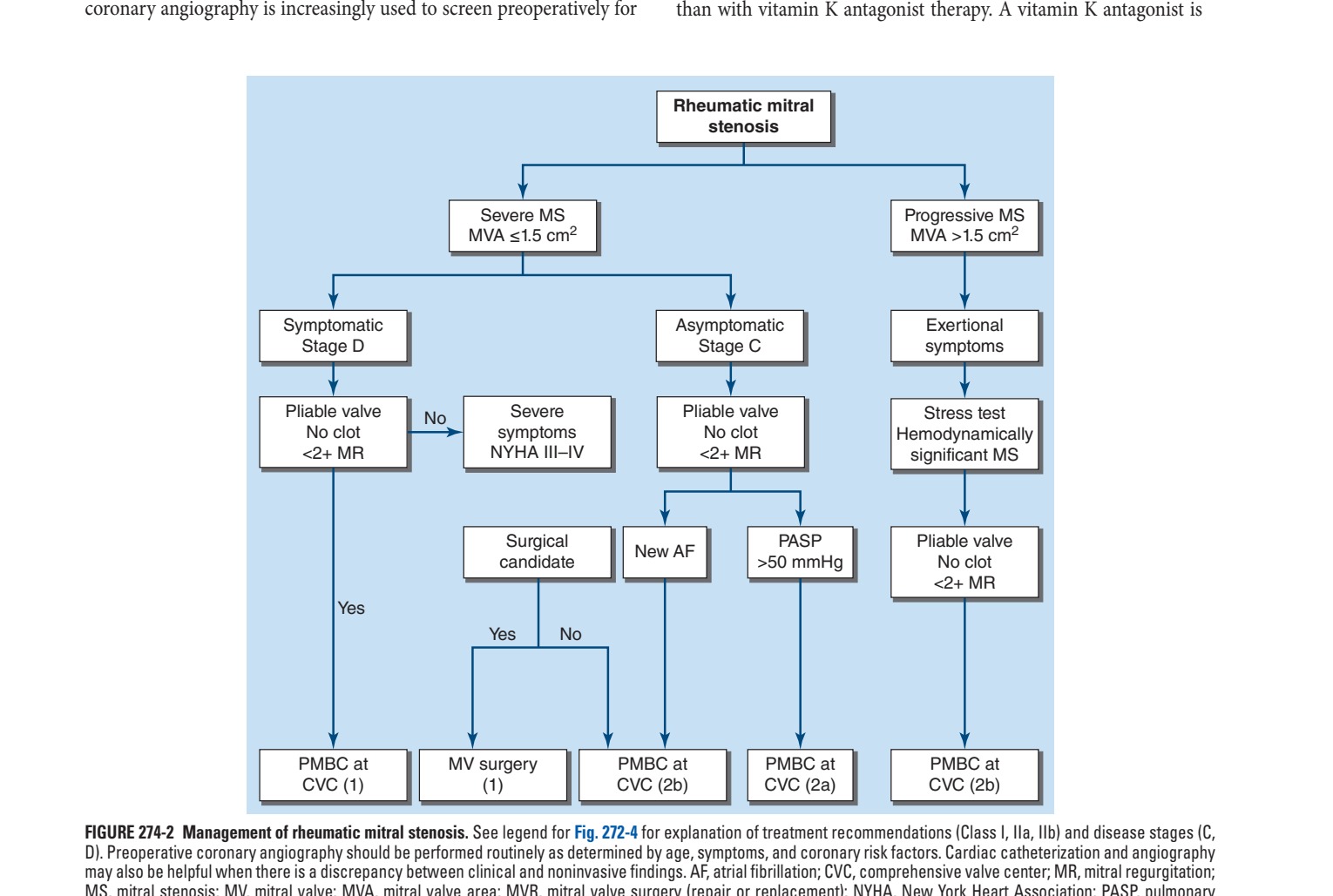
Caption: FIGURE 274-2 Management of rheumatic mitral stenosis. See legend for Fig. 272-4 for D). Preoperative coronary angiography should be performed routinely as determined by may also be helpful when there is a discrepancy between clinical and noninvasive MS, mitral stenosis; MV, mitral valve; MVA, mitral valve area; MVR, mitral valve arterial systolic pressure; PMBC, percutaneous mitral balloon commissurotomy. patients with valvular heart disease: A report of the American College of 143:e72, 2021.)
Flowchart 2
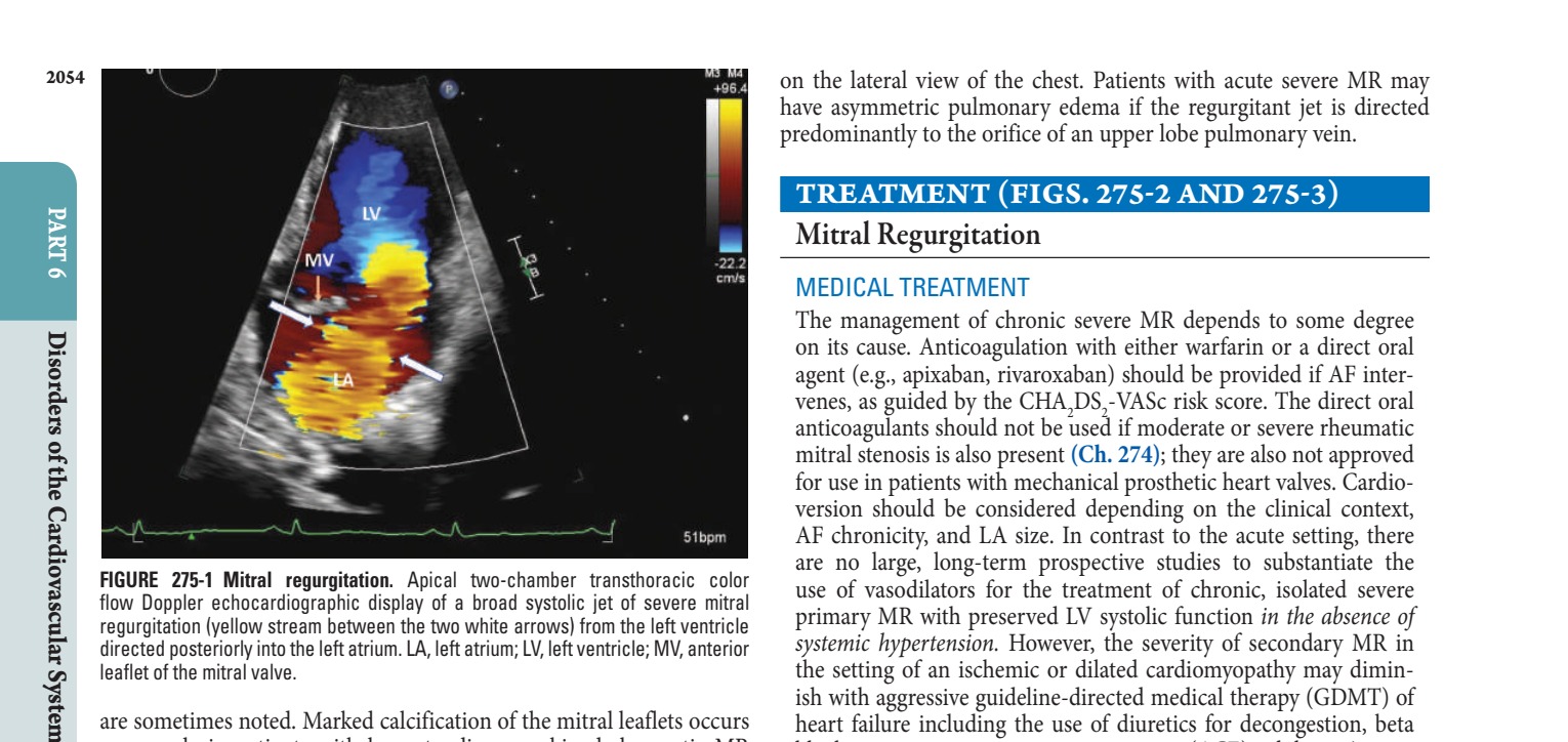
Caption: FIGURE 275-1 Mitral regurgitation. Apical two-chamber transthoracic color flow Doppler echocardiographic display of a broad systolic jet of severe mitral regurgitation (yellow stream between the two white arrows) from the left ventricle directed posteriorly into the left atrium. LA, left atrium; LV, left ventricle; MV, anterior leaflet of the mitral valve. are sometimes noted. Marked calcification of the mitral leaflets occurs commonly in patients with long-standing, combined rheumatic MR and MS, as well as in patients with radiation-induced mitral valve dis- ease. Calcification of the mitral annulus may be visualized, particularly
Flowchart 3
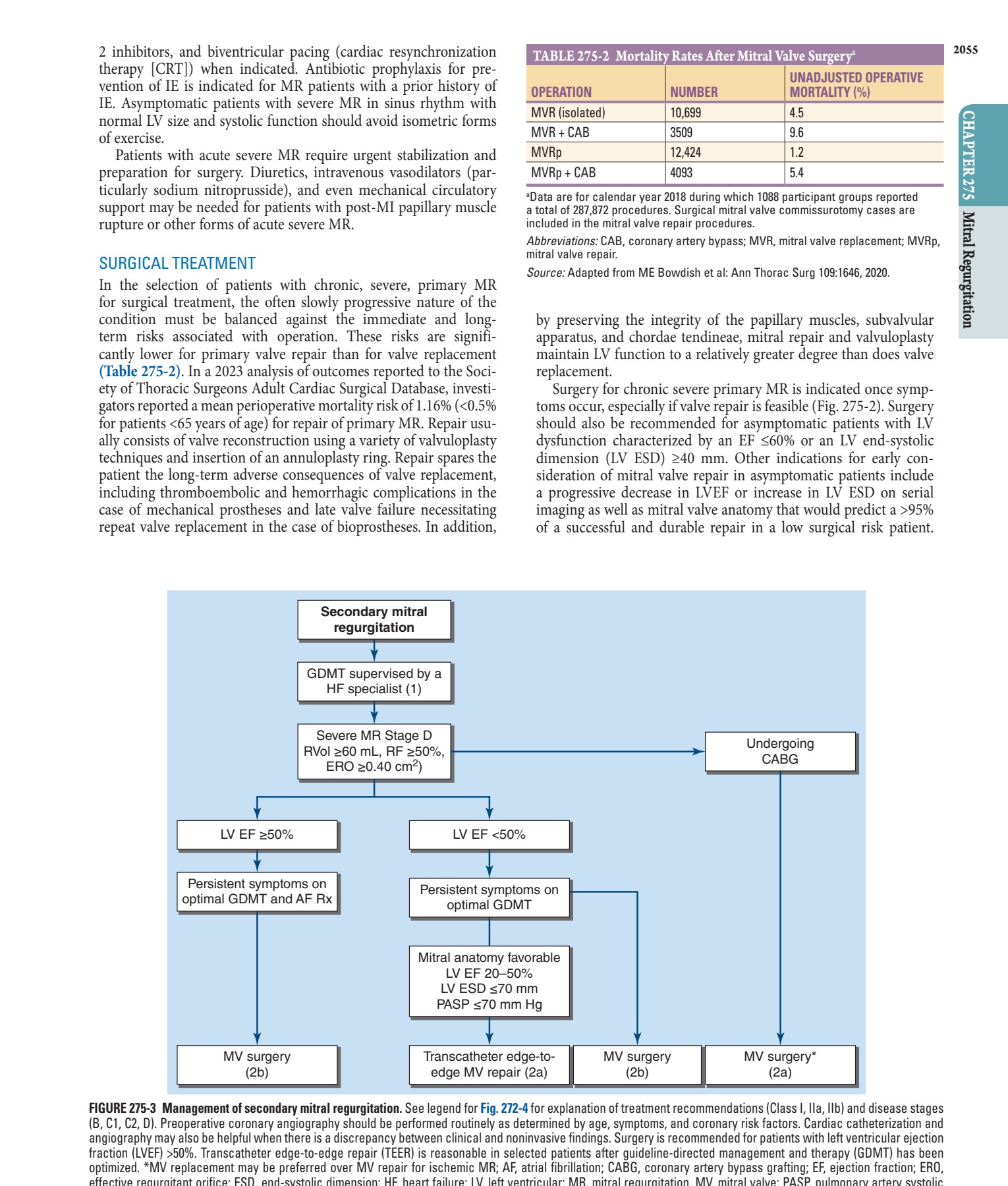
Caption: FIGURE 275-3 Management of secondary mitral regurgitation. See legend for Fig. 272-4 (B, C1, C2, D). Preoperative coronary angiography should be performed routinely as angiography may also be helpful when there is a discrepancy between clinical and fraction (LVEF) >50%. Transcatheter edge-to-edge repair (TEER) is reasonable in optimized. *MV replacement may be preferred over MV repair for ischemic MR; AF, effective regurgitant orifice; ESD, end-systolic dimension; HF, heart failure; LV, left pressure; RF, regurgitant fraction; RVol, regurgitant volume; Rx, treatment. of patients with valvular heart disease: A report of the American College of Circulation. 2021; 143(5):e72.)
Figures & Illustrations
Reproduced from Harrison's 22nd Edition.
Figure 1

Caption: FIGURE 275-4 Clip devices used to grasp the free edges of the anterior and posterior patients with mitral regurgitation. A. Fourth-generation of the first approved two paddles. The spacer is designed to reduce stress on the leaflets and preserve mitral is an Edwards Lifesciences tradename.) — Figure 274-1: Continuous wave Doppler interrogation of transmitral valve velocities in a patient with severe rheumatic mitral stenosis. Electrocardiogram on top. Vertical scale in meters/second. Horizontal scale in seconds at sweep speed of 75 mm/se. Velocity is converted to pressure using the Bernoulli equation. Mean mitral valve gradient calculated to 38 mmHg and mitral valve area to 0.7 cm2.
Figure 2

Caption: MVR is now routinely performed with preservation of the chordal FIGURE 274-3 Inoue balloon technique for percutaneous mitral balloon commissurotomy. A. After transseptal puncture, the deflated balloon catheter is attachments to optimize LV functional recovery. Perioperative mor- advanced across the interatrial septum, then across the mitral valve and into the tality rates with MVR vary with age, LV function, the presence of left ventricle. B–D. The balloon is inflated stepwise within the mitral orifice. CAD, and associated comorbidities. They average 2–5% overall but — Figure 274-2: Management of rheumatic mitral stenosis. Algorithm showing decision pathways for percutaneous mitral balloon commissurotomy (PMBC) versus surgical valve intervention (CVC) based on valve pliability, mitral valve area (MVA), presence of clot, NYHA functional class, and pulmonary arterial systolic pressure (PASP).
Figure 3

Caption: FIGURE 274-1 Continuous wave Doppler interrogation of transmitral valve velocities in a patient with severe rheumatic mitral stenosis. Electrocardiogram on top. Vertical scale in meters/second. Horizontal scale in seconds at sweep speed of 75 mm/se. Velocity is converted to pressure using the Bernoulli equation. In this example, the mean mitral valve gradient (area under the green tracing) is calculated to 38 mmHg and mitral valve area to 0.7 cm2. — Figure 274-3: Inoue balloon technique for percutaneous mitral balloon commissurotomy. A. After transseptal puncture, the deflated balloon catheter is advanced across the interatrial septum, then across the mitral valve and into the left ventricle. B–D. The balloon is inflated stepwise within the mitral orifice.
Figure 4

Caption: Figure 275-1: Mitral regurgitation. Apical two-chamber transthoracic color flow Doppler echocardiographic display of a broad systolic jet of severe mitral regurgitation (yellow stream between the two white arrows) from the left ventricle directed posteriorly into the left atrium.
Generated from Harrison's Principles of Internal Medicine, 22nd Edition.