Sleep Apnea
Chapter 308 | Part 7: Disorders of the Respiratory System · Part 7 – Respiratory Disorders
Detailed clinical reference synthesised from Harrison's Principles of Internal Medicine, 22nd Edition
🔑 Key Clinical Points
- OSA is defined by nocturnal/daytime symptoms plus sleep study findings (AHI ≥5 events/h) OR AHI ≥15 events/h without symptoms.
- CSA is less common, associated with heart failure, opioids, or high altitude, and presents with frequent awakenings and fatigue.
- CPAP is the standard first-line therapy; adherence is highly variable (average 50–80%) and improves with support.
- Oral appliances advance the mandible and work best for mild/moderate OSA or CPAP intolerance.
- ASV is contraindicated in patients with CSA and LVEF <45% due to increased mortality risk.
- Obesity is a major risk factor; a 10% weight gain is associated with a >30% increase in AHI.
- Hypopnea criteria: ≥30% airflow reduction for ≥10s with ≥3% desaturation OR arousal.
- Apnea criteria: Cessation of airflow for ≥10s with persistent effort (obstructive) or absence of effort (central).
- RERA: Partial obstruction with increasing inspiratory effort punctuated by arousal, without meeting hypopnea criteria.
- Women are often underdiagnosed due to lower arousal threshold and different presentation (REM sleep predominance).
- CPAP side effects include nasal congestion, claustrophobia, difficulty exhaling, and aerophagia; treatable with humidification or mask changes.
- Upper airway neurostimulation is an option for patients who fail CPAP, reducing AHI for at least 5 years.
- Vibrotactile positional therapy can reduce AHI by up to 50% in supine-predominant OSA.
- OSA is strongly associated with hypertension, cardiovascular disease, and metabolic disorders.
- AHI alone does not correlate strongly with degree of sleepiness; residual sleepiness may persist after treatment.
📑 Table of Contents
- 1. DEFINITION & OVERVIEW
- 1.1 Central Sleep Apnea
- 2. EPIDEMIOLOGY
- 2.1 Risk Factors
- 3. ETIOLOGY & PATHOPHYSIOLOGY
- 3.1 Anatomical Factors
- 3.2 Physiological Factors
- 4. CLINICAL FEATURES
- 4.1 Symptoms and History
- 4.2 Physical Findings
- 5. DIFFERENTIAL DIAGNOSIS
- 5.1 Central Sleep Apnea Differential
- 6. INVESTIGATIONS & DIAGNOSIS
- 6.1 Respiratory Event Definitions
- 6.2 Severity Scale
- 7. MANAGEMENT & TREATMENT
- 7.1 CPAP Therapy
- 7.2 Oral Appliances and Surgery
- 8. PROGNOSIS & COMPLICATIONS
- 8.1 Cardiovascular and Metabolic Risks
- 9. SPECIAL CONSIDERATIONS
- 9.1 Pediatric Considerations
- 9.2 Ethnic and Gender Considerations
- 10. KEY PEARLS & CLINICAL TRAPS
- 10.1 Diagnostic Clues
- 10.2 Exclusion Criteria
- Figures & Illustrations
📋 Figures in This Chapter
1. DEFINITION & OVERVIEW
- OSA and CSA are classified as sleep-related breathing disorders.
- OSA is the more common disorder, causing daytime sleepiness and impaired daily function.
- OSA is a cause of hypertension and is strongly associated with cardiovascular disease in adults and behavioral problems in children.
- CSA is less common and may occur alone or in combination with OSA.
- CSA can occur as a primary condition, as a response to high altitude, or secondary to a medical condition (e.g., heart failure) or medication (e.g., opioids).
- Patients with CSA often report frequent awakenings and daytime fatigue and are at increased risk for heart failure and atrial fibrillation.
- Chronic hyperventilation is a distinct condition that requires exclusion of acute hyperventilation causes (e.g., diabetic ketoacidosis, asthma) before diagnosis.
- Chronic hyperventilation is confirmed by arterial blood gas sampling demonstrating compensated respiratory alkalosis with near normal pH, low Paco2, and low calculated bicarbonate.
- A sustained 10% increase in alveolar ventilation is sufficient to perpetuate hypocapnia in chronic hyperventilation.
- OSA is defined on the basis of nocturnal and daytime symptoms as well as sleep study findings.
- Diagnosis requires the patient to have (1) either symptoms of nocturnal breathing disturbances (snoring, snorting, gasping, or breathing pauses during sleep) that impair quality of life or daytime sleepiness or fatigue that occurs despite sufficient opportunity to sleep and is unexplained by other medical problems; and (2) five or more episodes of obstructive apnea or hypopnea per hour of sleep (the apnea-hypopnea index [AHI], calculated as the number of episodes divided by the number of hours of sleep) documented during a sleep study.
- OSA also may be diagnosed in the absence of symptoms if the AHI is ≥15 episodes/h.
- Each episode of apnea or hypopnea represents a reduction in breathing for at least 10 s and commonly results in a ≥3% drop in oxygen saturation or a brain cortical arousal.
- OSA severity can be characterized by the frequency of breathing disturbances (AHI), the amount of oxyhemoglobin desaturation with respiratory events, the duration of apneas and hypopneas, the degree of sleep fragmentation, and the level of reported daytime sleepiness or functional impairment.
1.1 Central Sleep Apnea
- CSA is less common than OSA.
- CSA may occur in isolation or, more often, in combination with obstructive events in the form of mixed apneas.
- CSA is often caused by an increased sensitivity to PCO2, which leads to an unstable breathing pattern that manifests as hyperventilation alternating with apnea.
- A prolonged circulation delay between the pulmonary capillaries and carotid chemoreceptors is also a contributing cause; thus, individuals with congestive heart failure are at risk for CSA.
- With prolonged circulation delay, there is a crescendo-decrescendo breathing pattern known as Cheyne-Stokes breathing.
- Other risk factors for CSA include opioid medications (which appear to have a dose-dependent effect on CSA) and hypoxia (e.g., breathing at high altitude).
- In some individuals, CPAP—particularly at high pressures—seems to induce central apnea; this condition is referred to as complex sleep apnea or treatment-emergent central sleep apnea.
- Rarely, CSA may be caused by blunted chemosensitivity due to congenital disorders (congenital central hypoventilation syndrome) or acquired factors.
- CSA is associated with increased risk for the development of both heart failure and atrial fibrillation.
- Treatment of CSA is difficult and depends on the underlying cause.
- Limited data suggest that supplemental oxygen can reduce the frequency of central apneas, particularly in patients with hypoxemia; however, the effectiveness of supplemental oxygen on clinical outcomes is unknown.
- In patients with CSA or Cheyne-Stokes breathing associated with heart failure, treatment is directed at optimizing therapy for heart failure.
- Device-based therapies that adjust pressure support on a breath-by-breath basis (adaptive servoventilation [ASV]) can be effective for regularizing breathing.
- Two large international trials showed that ASV resulted in no cardiovascular or mortality benefit.
- The first trial unexpectedly reported increased mortality, leading to an FDA warning to avoid ASV in patients with CSA who have a left ventricular ejection fraction <45%.
- The second trial tested an ASV device that delivered lower pressure and reported no evidence of harm along with small improvements in symptoms, raising the possibility that ASV may have a role as an adjunctive therapy for patients with heart failure and CSA.
- Moderate to severe CSA in adults can be treated with an FDA-approved transvenous phrenic nerve stimulator.
- A clinical trial testing this therapy in patients with CSA from various etiologies showed an ~50% reduction in AHI and a near elimination of central apneas, along with improvements in sleep quality and quality of life.
2. EPIDEMIOLOGY
- The prevalence of OSA is twofold higher among men than among women.
- Factors that predispose men to OSA include android pattern of obesity (resulting in upper-airway and abdominal fat deposition) and relatively greater pharyngeal length, which increases collapsibility.
- Premenopausal women are relatively protected from OSA by the influence of sex hormones on ventilatory drive.
- The decline in sex difference in older age reflects an increased OSA prevalence in women after menopause.
- The pathogenesis and presentation of OSA also differ in men and women: compared to men, women have a lower arousal threshold and less neuromuscular collapsibility.
- Women tend to have shorter duration of apneas and apneas that occur predominantly in REM sleep.
- OSA prevalence varies with age, from 5 to 15% among middle-aged adults to >20% among elderly individuals, although in a majority of affected adults, the disorder is undiagnosed.
- There is a peak due to lymphoid hypertrophy among children between the ages of 3 and 8 years; with airway growth and lymphoid tissue regression during later childhood, prevalence declines.
- Then, as obesity prevalence increases in adolescence and adulthood, OSA prevalence again increases.
- The prevalence of OSA is especially high among patients with certain medical conditions, including diabetes mellitus, hypertension, and atrial fibrillation.
- Individuals of East Asian ancestry appear to be at increased risk of OSA at relatively low levels of body mass index, reflecting the greater influence of craniofacial risk factors.
- In the United States, African Americans, especially children and young adults, are at higher risk for OSA than their white counterparts.
- Approximately 40–60% of cases of OSA are attributable to excess weight.
- Obesity predisposes to OSA through the narrowing effects of upper airway fat on the pharyngeal lumen.
- Obesity also reduces chest wall compliance and decreases lung volumes, resulting in a loss of caudal traction on upper airway structures.
- Obese individuals are at a fourfold or greater risk for OSA than their normal-weight counterparts, although the association of OSA with obesity is weaker in the elderly than in younger adults.
- A 10% weight gain is associated with a >30% increase in AHI.
- Even modest weight loss or weight gain can influence the risk and severity of OSA.
- However, the absence of obesity does not exclude this diagnosis.
- OSA has a strong genetic basis, as evidenced by its significant familial aggregation and heritability.
- For a first-degree relative of a patient with OSA, the odds of having OSA are approximately twofold higher than that of someone without an affected relative.
- Several genetic variants have been associated with prevalence of OSA or with related traits, such as the frequency of apneas and hypopneas, the duration of respiratory events, and degree of overnight levels of hypoxemia.
2.1 Risk Factors
- Major risk factors for OSA are obesity, male sex, and older age.
- Additional risk factors include mandibular retrognathia and micrognathia, a positive family history of OSA, sedentary lifestyle, genetic syndromes that reduce upper airway patency (e.g., Down syndrome, Treacher-Collins syndrome), adeno-tonsillar hypertrophy (especially in children), menopause (in women), and various endocrine syndromes (e.g., acromegaly, hypothyroidism).
- Craniofacial factors such as mandibular retroposition or micrognathia, reflecting genetic variation or developmental influences, also can reduce lumen dimensions.
- Variations in craniofacial morphology that reduce the size of the posterior airway space increase OSA risk.
- The contribution of skeletal structural features to OSA is most evident in nonobese patients.
- Identification of features such as retrognathia can influence therapeutic decision-making.
- A high degree of nasal resistance (e.g., due to nasal septal deviation or polyps) can contribute to airway collapse by reducing intraluminal pressure downstream in the pharynx.
- High-level nasal resistance also may trigger mouth opening during sleep, which breaks the seal between the tongue and the palate and allows the tongue to fall posteriorly and occlude the airway.
3. ETIOLOGY & PATHOPHYSIOLOGY
- During inspiration, intraluminal pharyngeal pressure becomes increasingly negative, creating a suctioning force.
- Because the pharyngeal airway has no fixed bone or cartilage, airway patency is dependent on the stabilizing influence of the pharyngeal dilator muscles.
- Although these muscles are continuously activated during wakefulness, neuromuscular output declines with sleep onset.
- In patients with a collapsible airway, the reduction in neuromuscular output results in transient episodes of pharyngeal collapse (manifesting as an apnea) or near collapse (manifesting as a hypopnea).
- The episodes of collapse are typically terminated when ventilatory reflexes are activated and cause arousal, thus stimulating an increase in neuromuscular activity and opening of the airway.
- The airway may collapse at different sites, such as the soft palate (most common), tongue base, lateral pharyngeal walls, and/or epiglottis.
- OSA may be most severe during rapid eye movement (REM) sleep, when neuromuscular output to the skeletal muscles is particularly low, and in the supine position due to gravitational forces.
- Individuals with a small pharyngeal lumen require relatively high levels of neuromuscular activation to maintain patency during wakefulness and thus are predisposed to airway collapse following the normal sleep-related reduction in pharyngeal muscle activity during sleep.
- The airway lumen may be narrowed by enlargement of soft tissue structures (tongue, palate, and uvula) due to fat deposition, increased lymphoid tissue, or genetic variation.
- Low lung volume in the recumbent position, which is particularly pronounced in the obese, contributes to collapse (less caudal traction).
- Pharyngeal muscle activation is integrally linked to ventilatory drive.
- Thus, factors related to ventilatory control, particularly ventilatory sensitivity, arousal threshold, and neuromuscular responses to carbon dioxide (CO2), contribute to the pathogenesis of OSA.
- A buildup of CO2 during sleep activates both the diaphragm and the pharyngeal muscles.
- Pharyngeal activation stiffens the upper airway and can counteract inspiratory suction pressure and maintain airway patency to an extent that depends on the anatomic predisposition to collapse.
- Pharyngeal collapse can occur when the ventilatory control system is overly sensitive to CO2, with resultant wide fluctuations in ventilation, ventilatory drive, and upper airway stiffness.
- Moreover, pharyngeal collapse can occur when the ventilatory control system is overly sensitive to CO2, with resultant wide fluctuations in ventilation, ventilatory drive, and upper airway stiffness.
- Increasing levels of CO2 during sleep result in central nervous system arousal, causing the individual to move from a deeper to a lighter level of sleep or to awaken.
- A low arousal threshold (i.e., awakening to a low level of CO2 or ventilatory drive) can preempt the CO2-mediated process of pharyngeal muscle compensation and prevent airway stabilization.
- A high arousal threshold, conversely, may prevent appropriate termination of apneas, prolonging apnea duration and exacerbating oxyhemoglobin desaturation.
- Finally, any impairment in the ability of the muscles to compensate during sleep can contribute to collapse of the pharynx.
- The relative contributions of risk factors vary by age, sex, body mass index, and other factors.
- CSA pathophysiology involves an unstable breathing pattern that manifests as hyperventilation alternating with apnea.
- A prolonged circulation delay between the pulmonary capillaries and carotid chemoreceptors is also a contributing cause; thus, individuals with congestive heart failure are at risk for CSA.
- With prolonged circulation delay, there is a crescendo-decrescendo breathing pattern known as Cheyne-Stokes breathing.
- Other risk factors for CSA include opioid medications (which appear to have a dose-dependent effect on CSA) and hypoxia (e.g., breathing at high altitude).
- In some individuals, CPAP—particularly at high pressures—seems to induce central apnea; this condition is referred to as complex sleep apnea or treatment-emergent central sleep apnea.
- Rarely, CSA may be caused by blunted chemosensitivity due to congenital disorders (congenital central hypoventilation syndrome) or acquired factors.
3.1 Anatomical Factors
- The airway may collapse at different sites, such as the soft palate (most common), tongue base, lateral pharyngeal walls, and/or epiglottis.
- The airway lumen may be narrowed by enlargement of soft tissue structures (tongue, palate, and uvula) due to fat deposition, increased lymphoid tissue, or genetic variation.
- Craniofacial factors such as mandibular retroposition or micrognathia, reflecting genetic variation or developmental influences, also can reduce lumen dimensions.
- Variations in craniofacial morphology that reduce the size of the posterior airway space increase OSA risk.
- The contribution of skeletal structural features to OSA is most evident in nonobese patients.
- Identification of features such as retrognathia can influence therapeutic decision-making.
- A high degree of nasal resistance (e.g., due to nasal septal deviation or polyps) can contribute to airway collapse by reducing intraluminal pressure downstream in the pharynx.
- High-level nasal resistance also may trigger mouth opening during sleep, which breaks the seal between the tongue and the palate and allows the tongue to fall posteriorly and occlude the airway.
3.2 Physiological Factors
- Pharyngeal muscle activation is integrally linked to ventilatory drive.
- Thus, factors related to ventilatory control, particularly ventilatory sensitivity, arousal threshold, and neuromuscular responses to carbon dioxide (CO2), contribute to the pathogenesis of OSA.
- A buildup of CO2 during sleep activates both the diaphragm and the pharyngeal muscles.
- Pharyngeal activation stiffens the upper airway and can counteract inspiratory suction pressure and maintain airway patency to an extent that depends on the anatomic predisposition to collapse.
- Pharyngeal collapse can occur when the ventilatory control system is overly sensitive to CO2, with resultant wide fluctuations in ventilation, ventilatory drive, and upper airway stiffness.
- Increasing levels of CO2 during sleep result in central nervous system arousal, causing the individual to move from a deeper to a lighter level of sleep or to awaken.
- A low arousal threshold (i.e., awakening to a low level of CO2 or ventilatory drive) can preempt the CO2-mediated process of pharyngeal muscle compensation and prevent airway stabilization.
- A high arousal threshold, conversely, may prevent appropriate termination of apneas, prolonging apnea duration and exacerbating oxyhemoglobin desaturation.
- Finally, any impairment in the ability of the muscles to compensate during sleep can contribute to collapse of the pharynx.
- The relative contributions of risk factors vary by age, sex, body mass index, and other factors.
4. CLINICAL FEATURES
- Snoring is the most common symptom; however, its absence does not exclude the diagnosis, as pharyngeal collapse may occur without tissue vibration.
- Gasping or snorting during sleep may also be reported, reflecting termination of individual apneas with abrupt airway opening.
- Dyspnea is unusual, and its absence generally distinguishes OSA from paroxysmal nocturnal dyspnea, nocturnal asthma, and acid reflux with laryngospasm.
- Patients also may describe frequent awakening or sleep disruption, which is more common among women and older adults.
- The most common daytime symptom is excessive daytime sleepiness, identified by a history of difficulty maintaining alertness or involuntary periods of dozing.
- However, many women preferentially report fatigue rather than sleepiness.
- Other symptoms include a dry mouth, nocturnal heartburn, diaphoresis of the chest and neck, nocturia, morning headaches, trouble concentrating, irritability, and mood disturbances.
- Insomnia, which is common in the general population, may coexist with OSA.
- Although difficulty falling asleep is rarely caused by OSA, awakening at apnea termination may cause difficulty maintaining sleep, a symptom more likely to be reported by women than by men, and often responds to treatment in OSA.
- Physical findings often reflect the etiologic factors for the disorder as well as comorbid conditions, particularly vascular disease.
- On examination, patients may exhibit hypertension and regional (central) obesity, as indicated by a large waist and neck circumference.
- The oropharynx may reveal a small orifice with crowding due to an enlarged tongue, a low-lying soft palate with a bulky uvula, large tonsils, a high-arched palate, or micro-/retrognathia.
- Since nasal resistance can increase the propensity to pharyngeal collapse, the nasal cavity should be inspected for polyps, septal deviation, allergic rhinitis, and other signs of obstruction.
- Because patients with heart failure are at increased risk for both OSA and CSA, a careful cardiac examination should be conducted to detect possible left- or right-sided cardiac dysfunction.
- Evidence of cor pulmonale suggests a comorbid cardiopulmonary condition; OSA alone is not thought to cause right-heart failure.
- A neurologic evaluation is needed to evaluate for conditions such as neuromuscular and cerebrovascular diseases, which increase OSA risk.
4.1 Symptoms and History
- When possible, a sleep history should be obtained with assistance from a bed partner or household member.
- Snoring is the most common symptom; however, its absence does not exclude the diagnosis, as pharyngeal collapse may occur without tissue vibration.
- Gasping or snorting during sleep may also be reported, reflecting termination of individual apneas with abrupt airway opening.
- Dyspnea is unusual, and its absence generally distinguishes OSA from paroxysmal nocturnal dyspnea, nocturnal asthma, and acid reflux with laryngospasm.
- Patients also may describe frequent awakening or sleep disruption, which is more common among women and older adults.
- The most common daytime symptom is excessive daytime sleepiness, identified by a history of difficulty maintaining alertness or involuntary periods of dozing.
- However, many women preferentially report fatigue rather than sleepiness.
- Other symptoms include a dry mouth, nocturnal heartburn, diaphoresis of the chest and neck, nocturia, morning headaches, trouble concentrating, irritability, and mood disturbances.
- Insomnia, which is common in the general population, may coexist with OSA.
- Although difficulty falling asleep is rarely caused by OSA, awakening at apnea termination may cause difficulty maintaining sleep, a symptom more likely to be reported by women than by men, and often responds to treatment in OSA.
4.2 Physical Findings
- Physical findings often reflect the etiologic factors for the disorder as well as comorbid conditions, particularly vascular disease.
- On examination, patients may exhibit hypertension and regional (central) obesity, as indicated by a large waist and neck circumference.
- The oropharynx may reveal a small orifice with crowding due to an enlarged tongue, a low-lying soft palate with a bulky uvula, large tonsils, a high-arched palate, or micro-/retrognathia.
- Since nasal resistance can increase the propensity to pharyngeal collapse, the nasal cavity should be inspected for polyps, septal deviation, allergic rhinitis, and other signs of obstruction.
- Because patients with heart failure are at increased risk for both OSA and CSA, a careful cardiac examination should be conducted to detect possible left- or right-sided cardiac dysfunction.
- Evidence of cor pulmonale suggests a comorbid cardiopulmonary condition; OSA alone is not thought to cause right-heart failure.
- A neurologic evaluation is needed to evaluate for conditions such as neuromuscular and cerebrovascular diseases, which increase OSA risk.
5. DIFFERENTIAL DIAGNOSIS
- OSA vs CSA: CSA is less common and may occur alone or in combination with OSA. CSA is often caused by an increased sensitivity to PCO2, which leads to an unstable breathing pattern that manifests as hyperventilation alternating with apnea.
- OSA vs Narcolepsy: A multiple sleep latency test or a maintenance of wakefulness test can be useful in quantifying sleepiness and helping to distinguish OSA from narcolepsy.
- OSA vs Hyperventilation: Respiratory symptoms associated with acute hyperventilation can be the initial manifestation of systemic illnesses such as diabetic ketoacidosis. Causes of acute hyperventilation need to be excluded before a diagnosis of chronic hyperventilation is considered.
- OSA vs Nocturnal Asthma: Dyspnea is unusual in OSA and its absence generally distinguishes OSA from paroxysmal nocturnal dyspnea, nocturnal asthma, and acid reflux with laryngospasm.
- OSA vs GERD: Acid reflux with laryngospasm can mimic OSA symptoms.
- OSA vs Heart Failure: Patients with heart failure are at increased risk for both OSA and CSA. CSA is often caused by an increased sensitivity to PCO2, which leads to an unstable breathing pattern that manifests as hyperventilation alternating with apnea.
- OSA vs Idiopathic Hypoventilation: Waking hypoxemia or hypercarbia suggests coexisting cardiopulmonary disease or hypoventilation syndromes.
5.1 Central Sleep Apnea Differential
- CSA is less common than OSA.
- CSA may occur in isolation or, more often, in combination with obstructive events in the form of mixed apneas.
- CSA is often caused by an increased sensitivity to PCO2, which leads to an unstable breathing pattern that manifests as hyperventilation alternating with apnea.
- A prolonged circulation delay between the pulmonary capillaries and carotid chemoreceptors is also a contributing cause; thus, individuals with congestive heart failure are at risk for CSA.
- With prolonged circulation delay, there is a crescendo-decrescendo breathing pattern known as Cheyne-Stokes breathing.
- Other risk factors for CSA include opioid medications (which appear to have a dose-dependent effect on CSA) and hypoxia (e.g., breathing at high altitude).
- In some individuals, CPAP—particularly at high pressures—seems to induce central apnea; this condition is referred to as complex sleep apnea or treatment-emergent central sleep apnea.
- Rarely, CSA may be caused by blunted chemosensitivity due to congenital disorders (congenital central hypoventilation syndrome) or acquired factors.
- CSA is associated with increased risk for the development of both heart failure and atrial fibrillation.
- Treatment of CSA is difficult and depends on the underlying cause.
- Limited data suggest that supplemental oxygen can reduce the frequency of central apneas, particularly in patients with hypoxemia; however, the effectiveness of supplemental oxygen on clinical outcomes is unknown.
- In patients with CSA or Cheyne-Stokes breathing associated with heart failure, treatment is directed at optimizing therapy for heart failure.
6. INVESTIGATIONS & DIAGNOSIS
- The gold standard for diagnosis of OSA is an overnight polysomnogram (PSG).
- A negative in-laboratory PSG usually rules out OSA.
- However, false-negative studies can result from night-to-night variation in OSA severity, particularly if there was insufficient REM sleep or less supine sleep during testing than is typical for the patient.
- Home sleep tests that record only respiratory and cardiac channels are commonly used as a cost-effective means for diagnosing OSA.
- However, a home study may yield a false-negative result if sleep time is not accurately estimated or in individuals experiencing hypopneas with arousals rather than oxyhemoglobin desaturation.
- Therefore, if there is a high prior probability of OSA, a negative home study should be followed by PSG.
- The key physiologic information collected during a sleep study for OSA assessment includes measurement of breathing (changes in airflow, respiratory excursion), oxygenation (hemoglobin oxygen saturation), body position, and cardiac rhythm.
- In addition, PSGs and some home sleep studies measure sleep continuity and sleep stages (by electroencephalography, chin electromyography, electrooculography, and actigraphy), leg movements, and snoring intensity.
- This information is used to quantify the frequency and subtypes of abnormal respiratory events during sleep as well as associated changes in oxygen hemoglobin saturation, arousals, and sleep stage distributions.
- A typical sleep study report provides quantitative data such as the AHI (number of apneas plus hypopneas per hour of sleep) and the profile of oxygen saturation over the night (mean, nadir, time at low levels).
- Reports may also include the respiratory disturbance index, which includes the number of respiratory effort–related arousals in addition to the AHI.
- In-laboratory PSG also quantifies sleep latency (time from lights off to first sleep onset), the frequency of periodic limb movements during sleep, sleep efficiency (percentage of time asleep relative to time in bed), arousal index (number of cortical arousals per hour of sleep), and time in each sleep stage.
- These metrics can further characterize the severity of OSA, which is associated with an increased arousal index, low sleep efficiency, and a reduction of time in deep (stage N3) and REM sleep and increase in light (stage N1) sleep.
- The detection of autonomic responses to apneas and hypopneas, such as surges in blood pressure, changes in heart rate, and abnormalities in cardiac rhythm, also provides relevant information on OSA severity.
- While the AHI is the chief disease-defining measurement derived from sleep studies, metrics that quantify respiratory event–related hypoxemia, heart rate response, and ventilatory reduction have been shown to predict adverse cardiovascular outcomes and mortality and may soon be incorporated into clinical decision-making.
- Various imaging studies, including cephalometric radiography, upper airway magnetic resonance imaging (MRI) and computed tomography (CT), and fiberoptic endoscopy, can be used to identify anatomic risk factors for OSA.
- While these may be useful for planning surgical interventions, they are not indicated in the routine evaluation of OSA.
- Cardiac testing may yield evidence of impaired systolic or diastolic ventricular function or abnormal cardiac structure.
- Overnight blood pressure monitoring often displays a nondipping pattern (absence of the typical 10% fall of blood pressure during sleep compared to wakefulness).
- Arterial blood gas measurements made during wakefulness are usually normal.
- Waking hypoxemia or hypercarbia suggests coexisting cardiopulmonary disease or hypoventilation syndromes.
- Patients with severe nocturnal hypoxemia may have elevated hemoglobin values.
- A multiple sleep latency test or a maintenance of wakefulness test can be useful in quantifying sleepiness and helping to distinguish OSA from narcolepsy.
6.1 Respiratory Event Definitions
- Apnea: Cessation of airflow for ≥10 s during sleep, accompanied by:
- Persistent respiratory effort (obstructive apneas), or
- Absence of respiratory effort (central apneas).
- Hypopnea: A ≥30% reduction in airflow for at least 10 s during sleep that is accompanied by either a ≥3% desaturation or an arousal.
- Respiratory effort–related arousal (RERA): Partial obstruction that does not meet the criteria for hypopnea but provides evidence of increasing inspiratory effort (usually through pleural pressure monitoring) punctuated by an arousal.
- Flow-limited breath: A partially obstructed breath, typically within a hypopnea or RERA, identified by a flattened or scooped-out inspiratory flow shape.
Table 1 — TABLE 308-1 Respiratory Event Definitions
| Event | Definition |
|---|---|
| Apnea | Cessation of airflow for ≥10 s during sleep, accompanied by: Persistent respiratory effort (obstructive apneas), or Absence of respiratory effort (central apneas). |
| Hypopnea | A ≥30% reduction in airflow for at least 10 s during sleep that is accompanied by either a ≥3% desaturation or an arousal. |
| RERA | Partial obstruction that does not meet the criteria for hypopnea but provides evidence of increasing inspiratory effort (usually through pleural pressure monitoring) punctuated by an arousal. |
| Flow-limited breath | A partially obstructed breath, typically within a hypopnea or RERA, identified by a flattened or scooped-out inspiratory flow shape. |
6.2 Severity Scale
- Apnea-hypopnea index (AHI): Number of apneas plus hypopneas per hour of sleep.
- Respiratory disturbance index (RDI): Number of apneas plus hypopneas plus RERAs per hour of sleep.
- Mild OSAHS: AHI of 5–14 events/h.
- Moderate OSAHS: AHI of 15–29 events/h.
- Severe OSAHS: AHI of ≥30 events/h.
- Each level of AHI can be further quantified by level of sleepiness and associated hypoxemia.
Table 2 — TABLE 308-2 Obstructive Sleep Apnea/Hypopnea Syndrome (OSAHS): Quantification and Severity Scale
| Index | Definition |
|---|---|
| Apnea-hypopnea index (AHI) | Number of apneas plus hypopneas per hour of sleep |
| Respiratory disturbance index (RDI) | Number of apneas plus hypopneas plus RERAs per hour of sleep |
| Mild OSAHS | AHI of 5–14 events/h |
| Moderate OSAHS | AHI of 15–29 events/h |
| Severe OSAHS | AHI of ≥30 events/h |
7. MANAGEMENT & TREATMENT
- A comprehensive approach to the management of OSA is needed to reduce risk factors and comorbidities.
- The clinician should seek to identify and address lifestyle and behavioral factors as well as comorbidities that may be exacerbating OSA.
- As appropriate, treatment should aim to reduce weight; optimize sleep duration (7–9 h per night); regulate sleep schedules (with similar bedtimes and wake times across the week); encourage the patient to avoid sleeping in the supine position; treat nasal allergies; increase physical activity; eliminate alcohol ingestion (which impairs pharyngeal muscle activity) within 3 h of bedtime; and minimize use of sedative-hypnotic medications.
- CPAP is the standard medical therapy with the highest level of evidence for efficacy.
- Delivered through a nasal or nasal-oral mask, CPAP works as a mechanical splint to hold the airway open, thus maintaining airway patency during sleep.
- An overnight CPAP titration study can determine the optimal pressure setting that reduces the number of apneas/hypopneas during sleep, improves gas exchange, and reduces arousals.
- However, the use of auto-titrating CPAP (APAP) devices used in home settings has eliminated the need for titration sleep studies in many patients.
- Rates of adherence to CPAP treatment are highly variable (average, 50–80%) and may be improved with support by a skilled health care team who can address side effects, help the patient problem solve, and provide motivational education.
- Online CPAP support tools can provide the patient personalized support and feedback.
- Despite the limitations of CPAP, controlled studies have demonstrated its beneficial effect on alertness, mood, quality of life, work-related productivity, blood pressure, and insulin sensitivity.
- Uncontrolled studies also indicate a favorable effect on cardiovascular outcomes, cardiac ejection fraction, atrial fibrillation recurrence, and mortality risk.
- Oral appliances for OSA work by advancing the mandible, thus opening the airway by repositioning the lower jaw and pulling the tongue forward.
- These devices generally work better when customized for patient use; maximal adaptation can take several weeks.
- Efficacy studies show that these devices can reduce the AHI by ≥50% in two-thirds of individuals, although these data are based largely on patients with mild OSA.
- Some patients with moderate or severe OSA respond to oral appliances as well, although no consistent predictors of success have been identified in these groups, and thus, follow-up sleep testing is recommended.
- Side effects of oral appliances include temporomandibular joint pain and tooth movement; thus, they require that the patient have adequate dental and periodontal structures.
- Oral appliances are most often used for treating patients with mild/moderate OSA or patients who do not tolerate CPAP.
- However, as some patients are more adherent to oral appliances than to CPAP, these devices are under investigation for treatment of more severe disease.
- In some patients, more consistent treatment of OSA may be obtained by alternating between CPAP and oral appliance therapy.
- Upper airway surgery for OSA is less efficacious than CPAP and is mostly reserved for the treatment of patients who snore, have mild OSA, or cannot tolerate CPAP.
- Uvulopalatopharyngoplasty (UPPP, removal of the uvula and the margin of the soft palate) is the most commonly performed surgery for OSA.
- However, results vary greatly, and as a standalone procedure, UPPP often has limited efficacy, particularly in severe OSA and in obese patients.
- Thus, palatal surgery is often combined with other procedures (multilevel surgery involving more than one pharyngeal site/structure) performed by an experienced surgeon, but the selection of patients is an important factor and relies on careful targeting of culprit areas for surgical resection.
- Bariatric surgery is an option for obese patients with OSA and can improve not only OSA but also other obesity-associated health conditions.
- Other procedures that can decrease snoring but have minimal effects on OSA include injection of a hardening agent to the soft palate (resulting in stiffening), radiofrequency ablation, laser-assisted uvulopalatoplasty, and palatal implants.
- Upper airway neurostimulation is a recently tested alternative treatment for OSA.
- Unilateral stimulation of the hypoglossal nerve through a surgically implanted device was shown to significantly decrease the AHI and improve a number of patient-reported outcomes, such as sleepiness and quality of life, for a duration of at least 5 years after treatment in carefully selected patients.
- This therapy is reserved for patients who cannot tolerate or fail CPAP therapy.
- Current inclusion criteria are moderate to severe OSA (AHI 15–65), body mass index <35 kg/m2, and absence of complete concentric collapse at the level of the velum documented by awake and drug-induced endoscopy (a predictor of response to surgery).
- This therapy is also approved for use in children ages 13 or older and adults with Down’s syndrome who have an AHI of 10–50.
- Additional research is underway to further evaluate longer-term effectiveness and potential utility of this treatment in other patient groups.
- Vibrotactile positional therapy (devices that emit incremental vibratory stimuli in response to body position in the supine position) can reduce AHI by as much as 50% in individuals with OSA occurring predominantly in the supine position.
- Supplemental oxygen can improve oxygen saturation, but there is little evidence that it improves OSA symptoms or the AHI in unselected patients.
- There is conflicting evidence regarding the effect of supplemental oxygen on blood pressure in patients with OSA.
- Orofacial myofunctional therapy (OMT), which involves exercises designed to enhance the strength and coordination of orofacial muscles, has been tested in several small studies of OSA.
- These studies predominantly focused on individuals suffering from mild-to-moderate OSA and showed modest improvements in snoring and AHI, as well as in overall sleep quality.
- However, larger and more comprehensive studies are necessary to evaluate fully the efficacy of myofunctional therapy in treating OSA.
- Currently, there are no U.S. Food and Drug Administration (FDA)-approved medications for the treatment of OSA.
- However, ongoing research is exploring the efficacy of weight loss medications and drugs that enhance pharyngeal muscle activity during sleep.
- Among weight loss drugs, anorectics are generally not favored for OSA due to their side effects and potential for abuse.
- However, glucagon-like peptide 1 (GLP-1) receptor agonists have garnered recent interest because of their substantial impact on weight loss.
- Given the strong correlation between weight and OSA, drugs that facilitate weight reduction, such as GLP-1 agonists, which slow gastric emptying and suppress appetite, are being investigated for their potential in improving OSA symptoms.
- Liraglutide, a GLP-1 agonist, has shown small effects on OSA severity, but tirzepatide, which targets both GLP-1 and glucose-dependent insulinotropic polypeptide (GIP) receptors, may offer more significant weight loss and is currently under trial for OSA.
- Sodium-glucose cotransporter 2 (SGLT-2) inhibitors also promote weight loss and are under investigation for the treatment of OSA.
- Concerning drugs that stimulate pharyngeal muscle activity, studies have shown that sleep-related withdrawal of norepinephrine from cranial motor neurons leads to pharyngeal hypotonia and consequent upper airway obstruction in patients with OSA.
- Pharyngeal hypotonia is further exacerbated by muscarinic inhibition during REM sleep.
- Based on these mechanisms, drugs with noradrenergic and antimuscarinic properties have been tested for their potential in OSA management.
- Early studies have reported notable reductions in AHI, and more advanced phase 3 trials of these drug combinations are in progress.
7.1 CPAP Therapy
- CPAP is the standard medical therapy with the highest level of evidence for efficacy.
- Delivered through a nasal or nasal-oral mask, CPAP works as a mechanical splint to hold the airway open, thus maintaining airway patency during sleep.
- An overnight CPAP titration study can determine the optimal pressure setting that reduces the number of apneas/hypopneas during sleep, improves gas exchange, and reduces arousals.
- However, the use of auto-titrating CPAP (APAP) devices used in home settings has eliminated the need for titration sleep studies in many patients.
- Rates of adherence to CPAP treatment are highly variable (average, 50–80%) and may be improved with support by a skilled health care team who can address side effects, help the patient problem solve, and provide motivational education.
- Online CPAP support tools can provide the patient personalized support and feedback.
- Despite the limitations of CPAP, controlled studies have demonstrated its beneficial effect on alertness, mood, quality of life, work-related productivity, blood pressure, and insulin sensitivity.
- Uncontrolled studies also indicate a favorable effect on cardiovascular outcomes, cardiac ejection fraction, atrial fibrillation recurrence, and mortality risk.
Table 3 — TABLE 308-3 Side Effects of Continuous Positive Airway Pressure (CPAP) and Their Treatments
| Side Effect | Treatment |
|---|---|
| Nasal congestion | Provide heated humidification, administer saline/steroid nasal sprays |
| Claustrophobia | Change mask interface (e.g., to nasal prongs), promote habituation (i.e., practice breathing on CPAP while awake) |
| Difficulty exhaling | Temporarily reduce pressure, provide bilevel positive airway pressure |
| Bruised nasal ridge | Change mask interface, provide protective padding |
| Aerophagia | Administer antacids |
7.2 Oral Appliances and Surgery
- Oral appliances for OSA work by advancing the mandible, thus opening the airway by repositioning the lower jaw and pulling the tongue forward.
- These devices generally work better when customized for patient use; maximal adaptation can take several weeks.
- Efficacy studies show that these devices can reduce the AHI by ≥50% in two-thirds of individuals, although these data are based largely on patients with mild OSA.
- Some patients with moderate or severe OSA respond to oral appliances as well, although no consistent predictors of success have been identified in these groups, and thus, follow-up sleep testing is recommended.
- Side effects of oral appliances include temporomandibular joint pain and tooth movement; thus, they require that the patient have adequate dental and periodontal structures.
- Oral appliances are most often used for treating patients with mild/moderate OSA or patients who do not tolerate CPAP.
- However, as some patients are more adherent to oral appliances than to CPAP, these devices are under investigation for treatment of more severe disease.
- In some patients, more consistent treatment of OSA may be obtained by alternating between CPAP and oral appliance therapy.
- Upper airway surgery for OSA is less efficacious than CPAP and is mostly reserved for the treatment of patients who snore, have mild OSA, or cannot tolerate CPAP.
- Uvulopalatopharyngoplasty (UPPP, removal of the uvula and the margin of the soft palate) is the most commonly performed surgery for OSA.
- However, results vary greatly, and as a standalone procedure, UPPP often has limited efficacy, particularly in severe OSA and in obese patients.
- Thus, palatal surgery is often combined with other procedures (multilevel surgery involving more than one pharyngeal site/structure) performed by an experienced surgeon, but the selection of patients is an important factor and relies on careful targeting of culprit areas for surgical resection.
- Bariatric surgery is an option for obese patients with OSA and can improve not only OSA but also other obesity-associated health conditions.
- Other procedures that can decrease snoring but have minimal effects on OSA include injection of a hardening agent to the soft palate (resulting in stiffening), radiofrequency ablation, laser-assisted uvulopalatoplasty, and palatal implants.
- Upper airway neurostimulation is a recently tested alternative treatment for OSA.
- Unilateral stimulation of the hypoglossal nerve through a surgically implanted device was shown to significantly decrease the AHI and improve a number of patient-reported outcomes, such as sleepiness and quality of life, for a duration of at least 5 years after treatment in carefully selected patients.
- This therapy is reserved for patients who cannot tolerate or fail CPAP therapy.
- Current inclusion criteria are moderate to severe OSA (AHI 15–65), body mass index <35 kg/m2, and absence of complete concentric collapse at the level of the velum documented by awake and drug-induced endoscopy (a predictor of response to surgery).
- This therapy is also approved for use in children ages 13 or older and adults with Down’s syndrome who have an AHI of 10–50.
- Additional research is underway to further evaluate longer-term effectiveness and potential utility of this treatment in other patient groups.
- Vibrotactile positional therapy (devices that emit incremental vibratory stimuli in response to body position in the supine position) can reduce AHI by as much as 50% in individuals with OSA occurring predominantly in the supine position.
8. PROGNOSIS & COMPLICATIONS
- OSA is the most common medical cause of daytime sleepiness and negatively influences quality of life.
- It is also strongly associated with cardiac, cerebrovascular, and metabolic disorders and with premature death and increased risk for certain cancers.
- This broad range of health effects is attributable to the impact of sleep fragmentation, cortical arousal, and intermittent hypoxemia and hypercapnia on vascular, cardiac, metabolic, and neurohumoral functions.
- OSA-related respiratory events stimulate sympathetic overactivity, leading to acute blood pressure surges during sleep and nocturnal as well as daytime hypertension.
- OSA-related hypoxemia also stimulates release of acute-phase proteins and reactive oxygen species that exacerbate insulin resistance and lipolysis and cause an augmented prothrombotic and proinflammatory state.
- Inspiratory effort against an occluded airway causes large intrathoracic negative pressure swings, altering cardiac preload and afterload and resulting in cardiac remodeling and reduced cardiac function.
- Hypoxemia and sympathetic-parasympathetic imbalance also may cause electrical remodeling of the heart and myocyte injury.
- OSA significantly increases the risk of coronary artery disease, heart failure with and without reduced ejection fraction, atrial and ventricular arrhythmias, atherosclerosis and coronary artery disease, stroke, and diabetes.
- Treatment of OSA has been shown to reduce several markers of cardiovascular risk and improve insulin resistance and, in uncontrolled studies, is associated with a decreased recurrence rate of atrial fibrillation.
- Large randomized clinical trials, however, have failed to demonstrate that OSA treatment with CPAP reduces cardiovascular and stroke event rates or prolongs survival.
- These outcomes may reflect exclusion from these trials of patients with excessive sleepiness, as there is evidence that sleepy patients may have the greatest OSA-related cardiovascular risk.
- Limited adherence to treatment among trial participants or the widespread use of other effective secondary prevention measures, such as beta blockade, antiplatelet agents, and lipid-lowering therapy, may also limit the impact of CPAP on cardiovascular risk.
- More than 50% of patients with moderate to severe OSA report daytime sleepiness.
- However, there is not a strong association between AHI level and degree of sleepiness.
- Patients with OSA symptoms have a twofold increased risk of occupational accidents.
- Individuals with elevated AHIs are involved in motor vehicle crashes approximately two to three times as often as persons with normal AHIs.
- Randomized controlled trials have shown that treatment of OSA with CPAP alleviates sleepiness as measured by either questionnaire or objective testing in patients with both mild and more severe disease.
- However, the degree of improvement varies widely.
- Residual sleepiness may be due to several factors, including suboptimal treatment adherence, insufficient sleep duration, other sleep disorders, or prior hypoxia-mediated damage in brain areas involved in alertness.
- Moreover, visceral adipose tissue, which is present in higher amounts in patients with OSA, releases somnogenic cytokines that may contribute to sleepiness.
- Thus, even after treatment, it is important to assess and monitor patients for residual sleepiness and to optimize treatment adherence, improve sleep patterns, and identify other disorders that may contribute to sleepiness.
- Careful and supervised use of alerting agents may be appropriate as adjunctive treatment in patients in whom sleepiness does not respond to CPAP alone.
- Reductions in health-related quality of life are common in patients with OSA, with the largest decrements observed in scales that measure physical functioning and energy levels.
- Work-related productivity also has been shown to improve in patients with moderate to severe OSA treated with CPAP.
- Numerous studies, including a large-scale trial of minimally symptomatic patients, have shown that treatment with CPAP can improve these patient-reported outcomes.
- Depressive symptoms, in particular somatic symptoms (irritability, fatigue, lack of energy), are commonly reported in OSA and improve with CPAP.
8.1 Cardiovascular and Metabolic Risks
- OSA is the most common medical cause of daytime sleepiness and negatively influences quality of life.
- It is also strongly associated with cardiac, cerebrovascular, and metabolic disorders and with premature death and increased risk for certain cancers.
- This broad range of health effects is attributable to the impact of sleep fragmentation, cortical arousal, and intermittent hypoxemia and hypercapnia on vascular, cardiac, metabolic, and neurohumoral functions.
- OSA-related respiratory events stimulate sympathetic overactivity, leading to acute blood pressure surges during sleep and nocturnal as well as daytime hypertension.
- OSA-related hypoxemia also stimulates release of acute-phase proteins and reactive oxygen species that exacerbate insulin resistance and lipolysis and cause an augmented prothrombotic and proinflammatory state.
- Inspiratory effort against an occluded airway causes large intrathoracic negative pressure swings, altering cardiac preload and afterload and resulting in cardiac remodeling and reduced cardiac function.
- Hypoxemia and sympathetic-parasympathetic imbalance also may cause electrical remodeling of the heart and myocyte injury.
- OSA significantly increases the risk of coronary artery disease, heart failure with and without reduced ejection fraction, atrial and ventricular arrhythmias, atherosclerosis and coronary artery disease, stroke, and diabetes.
- Treatment of OSA has been shown to reduce several markers of cardiovascular risk and improve insulin resistance and, in uncontrolled studies, is associated with a decreased recurrence rate of atrial fibrillation.
- Large randomized clinical trials, however, have failed to demonstrate that OSA treatment with CPAP reduces cardiovascular and stroke event rates or prolongs survival.
- These outcomes may reflect exclusion from these trials of patients with excessive sleepiness, as there is evidence that sleepy patients may have the greatest OSA-related cardiovascular risk.
- Limited adherence to treatment among trial participants or the widespread use of other effective secondary prevention measures, such as beta blockade, antiplatelet agents, and lipid-lowering therapy, may also limit the impact of CPAP on cardiovascular risk.
9. SPECIAL CONSIDERATIONS
- OSA prevalence varies with age, from 5 to 15% among middle-aged adults to >20% among elderly individuals, although in a majority of affected adults, the disorder is undiagnosed.
- There is a peak due to lymphoid hypertrophy among children between the ages of 3 and 8 years; with airway growth and lymphoid tissue regression during later childhood, prevalence declines.
- Then, as obesity prevalence increases in adolescence and adulthood, OSA prevalence again increases.
- The prevalence of OSA is especially high among patients with certain medical conditions, including diabetes mellitus, hypertension, and atrial fibrillation.
- Individuals of East Asian ancestry appear to be at increased risk of OSA at relatively low levels of body mass index, reflecting the greater influence of craniofacial risk factors.
- In the United States, African Americans, especially children and young adults, are at higher risk for OSA than their white counterparts.
- Women tend to have shorter duration of apneas and apneas that occur predominantly in REM sleep.
- Failure to recognize these differences can contribute to underrecognition of OSA in women.
- CSA is associated with increased risk for the development of both heart failure and atrial fibrillation.
- Treatment of CSA has not been shown to improve long-term health outcomes, so specific treatment is generally indicated only for treatment of symptomatic CSA.
- In patients with CSA or Cheyne-Stokes breathing associated with heart failure, treatment is directed at optimizing therapy for heart failure.
- Device-based therapies that adjust pressure support on a breath-by-breath basis (adaptive servoventilation [ASV]) can be effective for regularizing breathing.
- Two large international trials showed that ASV resulted in no cardiovascular or mortality benefit.
- The first trial unexpectedly reported increased mortality, leading to an FDA warning to avoid ASV in patients with CSA who have a left ventricular ejection fraction <45%.
- The second trial tested an ASV device that delivered lower pressure and reported no evidence of harm along with small improvements in symptoms, raising the possibility that ASV may have a role as an adjunctive therapy for patients with heart failure and CSA.
- Moderate to severe CSA in adults can be treated with an FDA-approved transvenous phrenic nerve stimulator.
- A clinical trial testing this therapy in patients with CSA from various etiologies showed an ~50% reduction in AHI and a near elimination of central apneas, along with improvements in sleep quality and quality of life.
- Ongoing research is addressing long-term safety and effectiveness, as well as subgroups who may benefit the most from this intervention.
- Bariatric surgery is an option for obese patients with OSA and can improve not only OSA but also other obesity-associated health conditions.
- Other procedures that can decrease snoring but have minimal effects on OSA include injection of a hardening agent to the soft palate (resulting in stiffening), radiofrequency ablation, laser-assisted uvulopalatoplasty, and palatal implants.
- Upper airway neurostimulation is a recently tested alternative treatment for OSA.
- Unilateral stimulation of the hypoglossal nerve through a surgically implanted device was shown to significantly decrease the AHI and improve a number of patient-reported outcomes, such as sleepiness and quality of life, for a duration of at least 5 years after treatment in carefully selected patients.
- This therapy is reserved for patients who cannot tolerate or fail CPAP therapy.
- Current inclusion criteria are moderate to severe OSA (AHI 15–65), body mass index <35 kg/m2, and absence of complete concentric collapse at the level of the velum documented by awake and drug-induced endoscopy (a predictor of response to surgery).
- This therapy is also approved for use in children ages 13 or older and adults with Down’s syndrome who have an AHI of 10–50.
- Additional research is underway to further evaluate longer-term effectiveness and potential utility of this treatment in other patient groups.
- Vibrotactile positional therapy (devices that emit incremental vibratory stimuli in response to body position in the supine position) can reduce AHI by as much as 50% in individuals with OSA occurring predominantly in the supine position.
- Supplemental oxygen can improve oxygen saturation, but there is little evidence that it improves OSA symptoms or the AHI in unselected patients.
- There is conflicting evidence regarding the effect of supplemental oxygen on blood pressure in patients with OSA.
- Orofacial myofunctional therapy (OMT), which involves exercises designed to enhance the strength and coordination of orofacial muscles, has been tested in several small studies of OSA.
- These studies predominantly focused on individuals suffering from mild-to-moderate OSA and showed modest improvements in snoring and AHI, as well as in overall sleep quality.
- However, larger and more comprehensive studies are necessary to evaluate fully the efficacy of myofunctional therapy in treating OSA.
- Currently, there are no U.S. Food and Drug Administration (FDA)-approved medications for the treatment of OSA.
- However, ongoing research is exploring the efficacy of weight loss medications and drugs that enhance pharyngeal muscle activity during sleep.
- Among weight loss drugs, anorectics are generally not favored for OSA due to their side effects and potential for abuse.
- However, glucagon-like peptide 1 (GLP-1) receptor agonists have garnered recent interest because of their substantial impact on weight loss.
- Given the strong correlation between weight and OSA, drugs that facilitate weight reduction, such as GLP-1 agonists, which slow gastric emptying and suppress appetite, are being investigated for their potential in improving OSA symptoms.
- Liraglutide, a GLP-1 agonist, has shown small effects on OSA severity, but tirzepatide, which targets both GLP-1 and glucose-dependent insulinotropic polypeptide (GIP) receptors, may offer more significant weight loss and is currently under trial for OSA.
- Sodium-glucose cotransporter 2 (SGLT-2) inhibitors also promote weight loss and are under investigation for the treatment of OSA.
- Concerning drugs that stimulate pharyngeal muscle activity, studies have shown that sleep-related withdrawal of norepinephrine from cranial motor neurons leads to pharyngeal hypotonia and consequent upper airway obstruction in patients with OSA.
- Pharyngeal hypotonia is further exacerbated by muscarinic inhibition during REM sleep.
- Based on these mechanisms, drugs with noradrenergic and antimuscarinic properties have been tested for their potential in OSA management.
- Early studies have reported notable reductions in AHI, and more advanced phase 3 trials of these drug combinations are in progress.
9.1 Pediatric Considerations
- There is a peak due to lymphoid hypertrophy among children between the ages of 3 and 8 years; with airway growth and lymphoid tissue regression during later childhood, prevalence declines.
- Adeno-tonsillar hypertrophy (especially in children) is a risk factor.
- Upper airway neurostimulation is also approved for use in children ages 13 or older and adults with Down’s syndrome who have an AHI of 10–50.
9.2 Ethnic and Gender Considerations
- The prevalence of OSA is twofold higher among men than among women.
- Factors that predispose men to OSA include android pattern of obesity (resulting in upper-airway and abdominal fat deposition) and relatively greater pharyngeal length, which increases collapsibility.
- Premenopausal women are relatively protected from OSA by the influence of sex hormones on ventilatory drive.
- The decline in sex difference in older age reflects an increased OSA prevalence in women after menopause.
- The pathogenesis and presentation of OSA also differ in men and women: compared to men, women have a lower arousal threshold and less neuromuscular collapsibility.
- Women tend to have shorter duration of apneas and apneas that occur predominantly in REM sleep.
- Failure to recognize these differences can contribute to underrecognition of OSA in women.
- Individuals of East Asian ancestry appear to be at increased risk of OSA at relatively low levels of body mass index, reflecting the greater influence of craniofacial risk factors.
- In the United States, African Americans, especially children and young adults, are at higher risk for OSA than their white counterparts.
10. KEY PEARLS & CLINICAL TRAPS
- A high index of suspicion is required as increased minute ventilation can be difficult to detect on some physical examination.
- Once chronic hyperventilation is established, a sustained 10% increase in alveolar ventilation is sufficient to perpetuate hypocapnia.
- This increase can be accomplished with subtle changes in the respiratory pattern, such as occasional sigh breaths or yawning 2–3 times per min.
- There are few well-controlled treatment studies of chronic hyperventilation owing to its diverse features and the lack of a universally accepted diagnostic process.
- Clinicians often spend considerable time identifying initiating factors, excluding alternative diagnoses, and discussing the patient’s concerns and fears.
- In some patients, reassurance and frank discussion about hyperventilation can be liberating.
- Identifying and eliminating habits that perpetuate hypocapnia, such as frequent yawning or sigh breathing, can be helpful.
- Some evidence suggests that breathing exercises and diaphragmatic retraining may be beneficial for some patients.
- The evidence for using medications to treat hyperventilation is scant.
- Beta blockers may be helpful in patients with sympathetically mediated symptoms such as palpitations and tremors.
- Women are often underdiagnosed due to lower arousal threshold and different presentation (REM sleep predominance).
- CPAP adherence is highly variable (average, 50–80%) and may be improved with support by a skilled health care team who can address side effects, help the patient problem solve, and provide motivational education.
- ASV is contraindicated in patients with CSA who have a left ventricular ejection fraction <45% due to increased mortality risk.
- AHI alone does not correlate strongly with degree of sleepiness; residual sleepiness may persist after treatment.
- OSA is the most common medical cause of daytime sleepiness and negatively influences quality of life.
- It is also strongly associated with cardiac, cerebrovascular, and metabolic disorders and with premature death and increased risk for certain cancers.
- This broad range of health effects is attributable to the impact of sleep fragmentation, cortical arousal, and intermittent hypoxemia and hypercapnia on vascular, cardiac, metabolic, and neurohumoral functions.
- OSA-related respiratory events stimulate sympathetic overactivity, leading to acute blood pressure surges during sleep and nocturnal as well as daytime hypertension.
- OSA-related hypoxemia also stimulates release of acute-phase proteins and reactive oxygen species that exacerbate insulin resistance and lipolysis and cause an augmented prothrombotic and proinflammatory state.
- Inspiratory effort against an occluded airway causes large intrathoracic negative pressure swings, altering cardiac preload and afterload and resulting in cardiac remodeling and reduced cardiac function.
- Hypoxemia and sympathetic-parasympathetic imbalance also may cause electrical remodeling of the heart and myocyte injury.
- OSA significantly increases the risk of coronary artery disease, heart failure with and without reduced ejection fraction, atrial and ventricular arrhythmias, atherosclerosis and coronary artery disease, stroke, and diabetes.
- Treatment of OSA has been shown to reduce several markers of cardiovascular risk and improve insulin resistance and, in uncontrolled studies, is associated with a decreased recurrence rate of atrial fibrillation.
- Large randomized clinical trials, however, have failed to demonstrate that OSA treatment with CPAP reduces cardiovascular and stroke event rates or prolongs survival.
- These outcomes may reflect exclusion from these trials of patients with excessive sleepiness, as there is evidence that sleepy patients may have the greatest OSA-related cardiovascular risk.
- Limited adherence to treatment among trial participants or the widespread use of other effective secondary prevention measures, such as beta blockade, antiplatelet agents, and lipid-lowering therapy, may also limit the impact of CPAP on cardiovascular risk.
- More than 50% of patients with moderate to severe OSA report daytime sleepiness.
- However, there is not a strong association between AHI level and degree of sleepiness.
- Patients with OSA symptoms have a twofold increased risk of occupational accidents.
- Individuals with elevated AHIs are involved in motor vehicle crashes approximately two to three times as often as persons with normal AHIs.
- Randomized controlled trials have shown that treatment of OSA with CPAP alleviates sleepiness as measured by either questionnaire or objective testing in patients with both mild and more severe disease.
- However, the degree of improvement varies widely.
- Residual sleepiness may be due to several factors, including suboptimal treatment adherence, insufficient sleep duration, other sleep disorders, or prior hypoxia-mediated damage in brain areas involved in alertness.
- Moreover, visceral adipose tissue, which is present in higher amounts in patients with OSA, releases somnogenic cytokines that may contribute to sleepiness.
- Thus, even after treatment, it is important to assess and monitor patients for residual sleepiness and to optimize treatment adherence, improve sleep patterns, and identify other disorders that may contribute to sleepiness.
- Careful and supervised use of alerting agents may be appropriate as adjunctive treatment in patients in whom sleepiness does not respond to CPAP alone.
- Reductions in health-related quality of life are common in patients with OSA, with the largest decrements observed in scales that measure physical functioning and energy levels.
- Work-related productivity also has been shown to improve in patients with moderate to severe OSA treated with CPAP.
- Numerous studies, including a large-scale trial of minimally symptomatic patients, have shown that treatment with CPAP can improve these patient-reported outcomes.
- Depressive symptoms, in particular somatic symptoms (irritability, fatigue, lack of energy), are commonly reported in OSA and improve with CPAP.
10.1 Diagnostic Clues
- Snoring is the most common symptom; however, its absence does not exclude the diagnosis, as pharyngeal collapse may occur without tissue vibration.
- Gasping or snorting during sleep may also be reported, reflecting termination of individual apneas with abrupt airway opening.
- Dyspnea is unusual, and its absence generally distinguishes OSA from paroxysmal nocturnal dyspnea, nocturnal asthma, and acid reflux with laryngospasm.
- Patients also may describe frequent awakening or sleep disruption, which is more common among women and older adults.
- The most common daytime symptom is excessive daytime sleepiness, identified by a history of difficulty maintaining alertness or involuntary periods of dozing.
- However, many women preferentially report fatigue rather than sleepiness.
- Other symptoms include a dry mouth, nocturnal heartburn, diaphoresis of the chest and neck, nocturia, morning headaches, trouble concentrating, irritability, and mood disturbances.
- Insomnia, which is common in the general population, may coexist with OSA.
- Although difficulty falling asleep is rarely caused by OSA, awakening at apnea termination may cause difficulty maintaining sleep, a symptom more likely to be reported by women than by men, and often responds to treatment in OSA.
- Physical findings often reflect the etiologic factors for the disorder as well as comorbid conditions, particularly vascular disease.
- On examination, patients may exhibit hypertension and regional (central) obesity, as indicated by a large waist and neck circumference.
- The oropharynx may reveal a small orifice with crowding due to an enlarged tongue, a low-lying soft palate with a bulky uvula, large tonsils, a high-arched palate, or micro-/retrognathia.
- Since nasal resistance can increase the propensity to pharyngeal collapse, the nasal cavity should be inspected for polyps, septal deviation, allergic rhinitis, and other signs of obstruction.
- Because patients with heart failure are at increased risk for both OSA and CSA, a careful cardiac examination should be conducted to detect possible left- or right-sided cardiac dysfunction.
- Evidence of cor pulmonale suggests a comorbid cardiopulmonary condition; OSA alone is not thought to cause right-heart failure.
- A neurologic evaluation is needed to evaluate for conditions such as neuromuscular and cerebrovascular diseases, which increase OSA risk.
10.2 Exclusion Criteria
- A negative in-laboratory PSG usually rules out OSA.
- However, false-negative studies can result from night-to-night variation in OSA severity, particularly if there was insufficient REM sleep or less supine sleep during testing than is typical for the patient.
- Home sleep tests that record only respiratory and cardiac channels are commonly used as a cost-effective means for diagnosing OSA.
- However, a home study may yield a false-negative result if sleep time is not accurately estimated or in individuals experiencing hypopneas with arousals rather than oxyhemoglobin desaturation.
- Therefore, if there is a high prior probability of OSA, a negative home study should be followed by PSG.
- Waking hypoxemia or hypercarbia suggests coexisting cardiopulmonary disease or hypoventilation syndromes.
- Patients with severe nocturnal hypoxemia may have elevated hemoglobin values.
- A multiple sleep latency test or a maintenance of wakefulness test can be useful in quantifying sleepiness and helping to distinguish OSA from narcolepsy.
Figures & Illustrations
Reproduced from Harrison's 22nd Edition.
Figure 1

Caption: FIGURE 308-1 The structures causing airway collapse in obstructive sleep apnea include the palate, the tongue, and/or the epiglottis. In addition, collapse can also occur due to the lateral pharyngeal walls. — FIGURE 308-1: Anatomical structures causing airway collapse in obstructive sleep apnea, including the palate, tongue, epiglottis, and lateral pharyngeal walls.
Figure 2
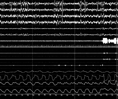
Caption: • Moderate OSAHS: AHI of 15–29 events/h FIGURE 308-3 Example of flow limitation. The inspiratory flow pattern in a patent • Severe OSAHS: AHI of ≥30 events/h airway is rounded and peaks in the middle. In contrast, a partially obstructed airway exhibits an early peak followed by mid-inspiratory flattening, yielding a scooped-out aEach level of AHI can be further quantified by level of sleepiness and associated appearance. hypoxemia. Abbreviation: RERAs, respiratory effort–related arousals. negative pressure swings, altering cardiac preload and afterload and Health Consequences and Comorbidities OSA is the most resulting in cardiac remodeling and reduced cardiac function. Hypox- common medical cause of daytime sleepiness and negatively influences emia and sympathetic-parasympathetic imbalance also may cause quality of life. It is also strongly associated with cardiac, cerebrovascu- — FIGURE 308-2A: Obstructive apnea showing cessation of airflow with persistent respiratory effort and chest-abdomen paradox on polysomnography.
Figure 3

Caption: • Moderate OSAHS: AHI of 15–29 events/h FIGURE 308-3 Example of flow limitation. The inspiratory flow pattern in a patent • Severe OSAHS: AHI of ≥30 events/h airway is rounded and peaks in the middle. In contrast, a partially obstructed airway exhibits an early peak followed by mid-inspiratory flattening, yielding a scooped-out aEach level of AHI can be further quantified by level of sleepiness and associated appearance. hypoxemia. Abbreviation: RERAs, respiratory effort–related arousals. negative pressure swings, altering cardiac preload and afterload and Health Consequences and Comorbidities OSA is the most resulting in cardiac remodeling and reduced cardiac function. Hypox- common medical cause of daytime sleepiness and negatively influences emia and sympathetic-parasympathetic imbalance also may cause quality of life. It is also strongly associated with cardiac, cerebrovascu- — FIGURE 308-2B: Central apnea in a patient with Cheyne-Stokes respiration due to congestive heart failure, showing absence of inspiratory effort.
Figure 4

Caption: • Moderate OSAHS: AHI of 15–29 events/h FIGURE 308-3 Example of flow limitation. The inspiratory flow pattern in a patent • Severe OSAHS: AHI of ≥30 events/h airway is rounded and peaks in the middle. In contrast, a partially obstructed airway exhibits an early peak followed by mid-inspiratory flattening, yielding a scooped-out aEach level of AHI can be further quantified by level of sleepiness and associated appearance. hypoxemia. Abbreviation: RERAs, respiratory effort–related arousals. negative pressure swings, altering cardiac preload and afterload and Health Consequences and Comorbidities OSA is the most resulting in cardiac remodeling and reduced cardiac function. Hypox- common medical cause of daytime sleepiness and negatively influences emia and sympathetic-parasympathetic imbalance also may cause quality of life. It is also strongly associated with cardiac, cerebrovascu- — FIGURE 308-2C: Hypopnea showing partial obstruction of the pharyngeal airway leading to desaturation and arousal.
Figure 5

Caption: • Moderate OSAHS: AHI of 15–29 events/h FIGURE 308-3 Example of flow limitation. The inspiratory flow pattern in a patent • Severe OSAHS: AHI of ≥30 events/h airway is rounded and peaks in the middle. In contrast, a partially obstructed airway exhibits an early peak followed by mid-inspiratory flattening, yielding a scooped-out aEach level of AHI can be further quantified by level of sleepiness and associated appearance. hypoxemia. Abbreviation: RERAs, respiratory effort–related arousals. negative pressure swings, altering cardiac preload and afterload and Health Consequences and Comorbidities OSA is the most resulting in cardiac remodeling and reduced cardiac function. Hypox- common medical cause of daytime sleepiness and negatively influences emia and sympathetic-parasympathetic imbalance also may cause quality of life. It is also strongly associated with cardiac, cerebrovascu- — FIGURE 308-3: Example of flow limitation in a partially obstructed airway, identified by a flattened or scooped-out inspiratory flow shape.
Generated from Harrison's Principles of Internal Medicine, 22nd Edition.